 Movie
Movie Controller

[English] 日本語
 Yorodumi
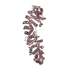
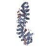
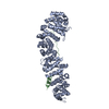
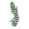
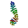
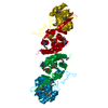
- PDB-1ee8: CRYSTAL STRUCTURE OF MUTM (FPG) PROTEIN FROM THERMUS THERMOPHILUS HB8 -
+ Open data

- Basic information
| Entry | Database: PDB / ID: 1ee8 | ||||||
|---|---|---|---|---|---|---|---|
| Title | CRYSTAL STRUCTURE OF MUTM (FPG) PROTEIN FROM THERMUS THERMOPHILUS HB8 | ||||||
 Components Components | MUTM (FPG) PROTEIN | ||||||
 Keywords Keywords | DNA BINDING PROTEIN / BETA SANDWICH / ZINC FINGER / HELIX TWO-TURNS HELIX / RIKEN Structural Genomics/Proteomics Initiative / RSGI / Structural Genomics | ||||||
| Function / homology |  Function and homology information Function and homology informationDNA-formamidopyrimidine glycosylase / 8-oxo-7,8-dihydroguanine DNA N-glycosylase activity / class I DNA-(apurinic or apyrimidinic site) endonuclease activity / DNA-(apurinic or apyrimidinic site) lyase / base-excision repair / damaged DNA binding / zinc ion binding Similarity search - Function | ||||||
| Biological species |   Thermus thermophilus (bacteria) Thermus thermophilus (bacteria) | ||||||
| Method |  X-RAY DIFFRACTION / SYNCHROTRON / Resolution: 1.9 Å X-RAY DIFFRACTION / SYNCHROTRON / Resolution: 1.9 Å | ||||||
 Authors Authors | Sugahara, M. / Mikawa, T. / Kumasaka, T. / Yamamoto, M. / Kato, R. / Fukuyama, K. / Inoue, Y. / Kuramitsu, S. / RIKEN Structural Genomics/Proteomics Initiative (RSGI) | ||||||
 Citation Citation | Journal: EMBO J. / Year: 2000 Title: Crystal structure of a repair enzyme of oxidatively damaged DNA, MutM (Fpg), from an extreme thermophile, Thermus thermophilus HB8. Authors: Sugahara, M. / Mikawa, T. / Kumasaka, T. / Yamamoto, M. / Kato, R. / Fukuyama, K. / Inoue, Y. / Kuramitsu, S. #1: Journal: Nucleic Acids Res. / Year: 1998Title: Termostable Repair Enzyme for Oxidative DNA Damage from Extremely Thermophilic Bacterium, Thermus Thermophilus HB8 Authors: Mikawa, T. / Kato, R. / Sugahara, M. / Kuramitsu, S. #2: Journal: J.BIOCHEM.(TOKYO) / Year: 2000Title: Crystallization and Preliminary X-ray Crystallographic Studies of Thermus Thermophilus HB8 MutM Protein Involved in Repairs Oxidative DNA Damage Authors: Sugahara, M. / Mikawa, T. / Kato, R. / Kumasaka, T. / Yamamoto, M. / Fukuyama, K. / Inoue, Y. / kuramitsu, S. | ||||||
| History |
|
- Structure visualization
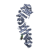
| Structure viewer | Molecule: MolmilJmol/JSmol |
|---|
- Downloads & links
-Download
| PDBx/mmCIF format | 1ee8.cif.gz | 119.4 KB | Display | PDBx/mmCIF format |
|---|---|---|---|---|
| PDB format | pdb1ee8.ent.gz | 93.3 KB | Display | PDB format |
| PDBx/mmJSON format | 1ee8.json.gz | Tree view | PDBx/mmJSON format | |
| Others |  Other downloads Other downloads |
-Validation report
| Arichive directory | https://data.pdbj.org/pub/pdb/validation_reports/ee/1ee8ftp://data.pdbj.org/pub/pdb/validation_reports/ee/1ee8 | HTTPS FTP |
|---|
-Related structure data
| Similar structure data | |
|---|---|
| Other databases |
-Links
 PDBj
PDBj


- Assembly
| Deposited unit | 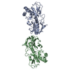
| ||||||||
|---|---|---|---|---|---|---|---|---|---|
| 1 |
| ||||||||
| Unit cell |
|
-Components
| #1: Protein | Mass: 29833.305 Da / Num. of mol.: 2 Source method: isolated from a genetically manipulated source Source: (gene. exp.) Thermus thermophilus (bacteria) / Strain: HB8 / Plasmid: PET3A / Species (production host): Escherichia coli / Production host: #2: Chemical |   Mass: 65.409 Da / Num. of mol.: 2 / Source method: obtained synthetically / Formula: Zn Mass: 65.409 Da / Num. of mol.: 2 / Source method: obtained synthetically / Formula: Zn#3: Water | ChemComp-HOH / |  Mass: 18.015 Da / Num. of mol.: 292 / Source method: isolated from a natural source / Formula: H2O Mass: 18.015 Da / Num. of mol.: 292 / Source method: isolated from a natural source / Formula: H2O |
|---|
-Experimental details
-Experiment
| Experiment | Method: X-RAY DIFFRACTION / Number of used crystals: 1 |
|---|
- Sample preparation
| Crystal | Density Matthews: 2.24 Å3/Da / Density % sol: 45.11 % |
|---|---|
| Crystal grow | Temperature: 293 K / Method: vapor diffusion, hanging drop / pH: 9.8 Details: PEG MME 2000, 2-mercaptoethanol, CHES, pH 9.8, VAPOR DIFFUSION, HANGING DROP, temperature 293.0K |
| Crystal grow | *PLUS Details: Sugahara, M., (2000) J. Biochem., 127, 9. |
-Data collection
| Diffraction | Mean temperature: 100 K |
|---|---|
| Diffraction source | Source: SYNCHROTRON / Site: SPring-8  / Beamline: BL45XU / Wavelength: 1.5418 / Beamline: BL45XU / Wavelength: 1.5418 |
| Radiation | Protocol: SINGLE WAVELENGTH / Monochromatic (M) / Laue (L): M / Scattering type: x-ray |
| Radiation wavelength | Wavelength: 1.5418 Å / Relative weight: 1 |
| Reflection | Resolution: 1.9→50 Å / Num. all: 37892 / Num. obs: 37892 / % possible obs: 0.9 % / Observed criterion σ(F): -3 / Observed criterion σ(I): -3 / Redundancy: 4.4 % / Biso Wilson estimate: 25.65 Å2 / Rmerge(I) obs: 0.063 / Net I/σ(I): 29.1 |
| Reflection shell | Resolution: 1.9→1.93 Å / Redundancy: 3.96 % / Rmerge(I) obs: 0.91 / Num. unique all: 1730 / % possible all: 0.821 |
| Reflection | *PLUS % possible obs: 90 % / Num. measured all: 402397 / Rmerge(I) obs: 0.07 |
| Reflection shell | *PLUS % possible obs: 82.1 % / Num. unique obs: 1729 / Rmerge(I) obs: 0.62 / Mean I/σ(I) obs: 3.2 |
- Processing
| Software |
| ||||||||||||||||||||||||||||||||||||
|---|---|---|---|---|---|---|---|---|---|---|---|---|---|---|---|---|---|---|---|---|---|---|---|---|---|---|---|---|---|---|---|---|---|---|---|---|---|
| Refinement | Resolution: 1.9→50 Å / Cross valid method: THROUGHOUT / σ(F): 2 / σ(I): 2
| ||||||||||||||||||||||||||||||||||||
| Refinement step | Cycle: LAST / Resolution: 1.9→50 Å
| ||||||||||||||||||||||||||||||||||||
| Software | *PLUS Name: CNS / Version: 0.9 / Classification: refinement | ||||||||||||||||||||||||||||||||||||
| Refinement | *PLUS Highest resolution: 1.9 Å / Lowest resolution: 50 Å / σ(F): 2 / % reflection Rfree: 10 % / Rfactor obs: 0.214 | ||||||||||||||||||||||||||||||||||||
| Solvent computation | *PLUS | ||||||||||||||||||||||||||||||||||||
| Displacement parameters | *PLUS Biso mean: 30.82 Å2 | ||||||||||||||||||||||||||||||||||||
| Refine LS restraints | *PLUS
|
