 Movie
Movie Controller

[English] 日本語
 Yorodumi
- PDB-3a7q: Structural basis for specific recognition of reelin by its receptors -
+ Open data

- Basic information
| Entry | Database: PDB / ID: 3a7q | |||||||||
|---|---|---|---|---|---|---|---|---|---|---|
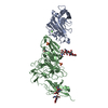
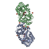
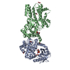
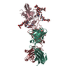
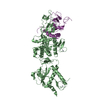
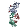
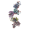
| Title | Structural basis for specific recognition of reelin by its receptors | |||||||||
 Components Components |
| |||||||||
 Keywords Keywords | SIGNALING PROTEIN | |||||||||
| Function / homology |  Function and homology information Function and homology informationammon gyrus development / spinal cord patterning / cerebral cortex tangential migration / reelin complex / positive regulation of lateral motor column neuron migration / lateral motor column neuron migration / Reelin signalling pathway / lipoprotein particle receptor binding / regulation of synaptic activity / reelin receptor activity ...ammon gyrus development / spinal cord patterning / cerebral cortex tangential migration / reelin complex / positive regulation of lateral motor column neuron migration / lateral motor column neuron migration / Reelin signalling pathway / lipoprotein particle receptor binding / regulation of synaptic activity / reelin receptor activity / postsynaptic density assembly / postsynaptic density protein 95 clustering / layer formation in cerebral cortex / very-low-density lipoprotein particle receptor activity / positive regulation of synapse maturation / positive regulation of protein tyrosine kinase activity / ventral spinal cord development / regulation of behavior / receptor localization to synapse / NMDA glutamate receptor clustering / low-density lipoprotein particle receptor activity / regulation of neuron migration / glial cell differentiation / dentate gyrus development / very-low-density lipoprotein particle receptor binding / reelin-mediated signaling pathway / positive regulation of dendritic spine morphogenesis / positive regulation of AMPA receptor activity / protein localization to synapse / positive regulation of small GTPase mediated signal transduction / dendrite morphogenesis / regulation of synapse maturation / positive regulation of dendrite development / response to pain / forebrain development / regulation of NMDA receptor activity / regulation of neuron differentiation / cargo receptor activity / Platelet sensitization by LDL / regulation of innate immune response / dendrite development / positive regulation of protein kinase activity / associative learning / Hydrolases; Acting on peptide bonds (peptidases); Serine endopeptidases / apolipoprotein binding / synaptic vesicle exocytosis / positive regulation of TOR signaling / long-term memory / Retinoid metabolism and transport / positive regulation of synaptic transmission, glutamatergic / retinoid metabolic process / peptidyl-tyrosine phosphorylation / axon guidance / learning / serine-type peptidase activity / hippocampus development / positive regulation of excitatory postsynaptic potential / locomotory behavior / positive regulation of long-term synaptic potentiation / central nervous system development / lipid metabolic process / brain development / positive regulation of neuron projection development / neuron migration / cerebral cortex development / cell morphogenesis / positive regulation of protein phosphorylation / caveola / postsynaptic density membrane / modulation of chemical synaptic transmission / long-term synaptic potentiation / Schaffer collateral - CA1 synapse / cytokine-mediated signaling pathway / endocytosis / calcium-dependent protein binding / transmembrane signaling receptor activity / cell migration / amyloid-beta binding / presynapse / regulation of gene expression / extracellular matrix / chemical synaptic transmission / regulation of apoptotic process / perikaryon / positive regulation of phosphatidylinositol 3-kinase/protein kinase B signal transduction / signaling receptor complex / cell adhesion / neuron projection / receptor ligand activity / axon / neuronal cell body / calcium ion binding / dendrite / glutamatergic synapse / cell surface / signal transduction / proteolysis / : / membrane / metal ion binding Similarity search - Function | |||||||||
| Biological species |   Homo sapiens (human) Homo sapiens (human) | |||||||||
| Method |  X-RAY DIFFRACTION / SYNCHROTRON / MOLECULAR REPLACEMENT / Resolution: 2.6 Å X-RAY DIFFRACTION / SYNCHROTRON / MOLECULAR REPLACEMENT / Resolution: 2.6 Å | |||||||||
 Authors Authors | Yasui, N. / Nogi, T. / Takagi, J. | |||||||||
 Citation Citation | Journal: Structure / Year: 2010 Title: Structural Basis for Specific Recognition of Reelin by Its Receptors Authors: Yasui, N. / Nogi, T. / Takagi, J. | |||||||||
| History |
|
- Structure visualization
| Structure viewer | Molecule: MolmilJmol/JSmol |
|---|
- Downloads & links
-Download
| PDBx/mmCIF format | 3a7q.cif.gz | 162.1 KB | Display | PDBx/mmCIF format |
|---|---|---|---|---|
| PDB format | pdb3a7q.ent.gz | 122.6 KB | Display | PDB format |
| PDBx/mmJSON format | 3a7q.json.gz | Tree view | PDBx/mmJSON format | |
| Others |  Other downloads Other downloads |
-Validation report
| Arichive directory | https://data.pdbj.org/pub/pdb/validation_reports/a7/3a7qftp://data.pdbj.org/pub/pdb/validation_reports/a7/3a7q | HTTPS FTP |
|---|
-Related structure data
| Related structure data | 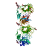 2e26S S: Starting model for refinement |
|---|---|
| Similar structure data |


-Links
 PDBj
PDBj


- Assembly
| Deposited unit | 
| ||||||||
|---|---|---|---|---|---|---|---|---|---|
| 1 |
| ||||||||
| 2 | 
| ||||||||
| 3 | 
| ||||||||
| Unit cell |
|
-Components
-Protein / Protein/peptide , 2 types, 2 molecules AB
| #1: Protein | Mass: 81317.078 Da / Num. of mol.: 1 / Fragment: repeat 5-6 fragment, UNP residues 1948-2661 / Mutation: C2101A Source method: isolated from a genetically manipulated source Source: (gene. exp.)  Cricetulus griseus (Chinese hamster) / Strain (production host): lec 3.2.8.1 / Tissue (production host): ovary Cricetulus griseus (Chinese hamster) / Strain (production host): lec 3.2.8.1 / Tissue (production host): ovaryReferences: UniProt: Q60841, Hydrolases; Acting on peptide bonds (peptidases); Serine endopeptidases |
|---|---|
| #2: Protein/peptide | Mass: 5082.448 Da / Num. of mol.: 1 / Fragment: LA1 module, UNP residues 42-83 Source method: isolated from a genetically manipulated source Source: (gene. exp.) Homo sapiens (human) / Gene: APOER2 / Plasmid: pGEX-3T / Production host:  |
-Sugars , 2 types, 4 molecules 
| #3: Polysaccharide | 2-acetamido-2-deoxy-beta-D-glucopyranose-(1-4)-2-acetamido-2-deoxy-beta-D-glucopyranose Source method: isolated from a genetically manipulated source |
|---|---|
| #4: Sugar |  Type: D-saccharide, beta linking / Mass: 221.208 Da / Num. of mol.: 3 Type: D-saccharide, beta linking / Mass: 221.208 Da / Num. of mol.: 3Source method: isolated from a genetically manipulated source Formula: C8H15NO6 |
-Non-polymers , 3 types, 25 molecules 


| #5: Chemical | ChemComp-CA /  Mass: 40.078 Da / Num. of mol.: 5 / Source method: obtained synthetically / Formula: Ca Mass: 40.078 Da / Num. of mol.: 5 / Source method: obtained synthetically / Formula: Ca#6: Chemical | ChemComp-ZN / |  Mass: 65.409 Da / Num. of mol.: 1 / Source method: obtained synthetically / Formula: Zn Mass: 65.409 Da / Num. of mol.: 1 / Source method: obtained synthetically / Formula: Zn#7: Water | ChemComp-HOH / | Mass: 18.015 Da / Num. of mol.: 19 / Source method: isolated from a natural source / Formula: H2O |
|---|
-Details
| Has protein modification | Y |
|---|---|
| Sequence details | THE SEQUENCE IS BASED ON REFERENCE 1, 2, 3, 5 AND 12 IN THE DATABASE UNIPROTKB/SWISS-PROT Q14114 ...THE SEQUENCE IS BASED ON REFERENCE 1, 2, 3, 5 AND 12 IN THE DATABASE UNIPROTKB/SWISS-PROT Q14114 (LRP8_HUMAN). D46E IS NATURAL VARIENT OF LRP8_HUMAN. |
-Experimental details
-Experiment
| Experiment | Method: X-RAY DIFFRACTION / Number of used crystals: 1 |
|---|
- Sample preparation
| Crystal | Density Matthews: 2.17 Å3/Da / Density % sol: 43.29 % |
|---|---|
| Crystal grow | Temperature: 293 K / Method: vapor diffusion, hanging drop / pH: 7.5 Details: 36-38% MPD, 21-25% PEG 1000, 100mM HEPES-Na pH7.5, VAPOR DIFFUSION, HANGING DROP, temperature 293K |
-Data collection
| Diffraction | Mean temperature: 100 K |
|---|---|
| Diffraction source | Source: SYNCHROTRON / Site: SPring-8  / Beamline: BL44XU / Wavelength: 0.9 Å / Beamline: BL44XU / Wavelength: 0.9 Å |
| Detector | Type: Bruker DIP-6040 / Detector: CCD / Date: Oct 3, 2006 |
| Radiation | Protocol: SINGLE WAVELENGTH / Monochromatic (M) / Laue (L): M / Scattering type: x-ray |
| Radiation wavelength | Wavelength: 0.9 Å / Relative weight: 1 |
| Reflection | Resolution: 2.6→46.93 Å / Num. obs: 21990 / % possible obs: 96.6 % / Observed criterion σ(I): -3 / Redundancy: 3.7 % / Biso Wilson estimate: 56 Å2 / Rmerge(I) obs: 0.074 / Rsym value: 0.074 / Net I/σ(I): 7.5 |
| Reflection shell | Resolution: 2.6→2.74 Å / Redundancy: 3.5 % / Rmerge(I) obs: 0.374 / Mean I/σ(I) obs: 1.8 / Num. unique all: 3213 / Rsym value: 0.374 / % possible all: 97.5 |
- Processing
| Software |
| ||||||||||||||||||||||||||||||||||||||||||||||||||||||||||||||||||||||||||||||||||||||||||||||||||||||||||||||||||||||||||||||||||||||||||||||||||||||||||||||||||||||||||||||||||||||||||||||||||||||||
|---|---|---|---|---|---|---|---|---|---|---|---|---|---|---|---|---|---|---|---|---|---|---|---|---|---|---|---|---|---|---|---|---|---|---|---|---|---|---|---|---|---|---|---|---|---|---|---|---|---|---|---|---|---|---|---|---|---|---|---|---|---|---|---|---|---|---|---|---|---|---|---|---|---|---|---|---|---|---|---|---|---|---|---|---|---|---|---|---|---|---|---|---|---|---|---|---|---|---|---|---|---|---|---|---|---|---|---|---|---|---|---|---|---|---|---|---|---|---|---|---|---|---|---|---|---|---|---|---|---|---|---|---|---|---|---|---|---|---|---|---|---|---|---|---|---|---|---|---|---|---|---|---|---|---|---|---|---|---|---|---|---|---|---|---|---|---|---|---|---|---|---|---|---|---|---|---|---|---|---|---|---|---|---|---|---|---|---|---|---|---|---|---|---|---|---|---|---|---|---|---|---|
| Refinement | Method to determine structure: MOLECULAR REPLACEMENT Starting model: PDB ENTRY 2E26 Resolution: 2.6→46.93 Å / Cor.coef. Fo:Fc: 0.924 / Cor.coef. Fo:Fc free: 0.866 / SU B: 30.323 / SU ML: 0.301 / TLS residual ADP flag: LIKELY RESIDUAL / Cross valid method: THROUGHOUT / ESU R Free: 0.418 / Stereochemistry target values: MAXIMUM LIKELIHOOD / Details: HYDROGENS HAVE BEEN ADDED IN THE RIDING POSITIONS
| ||||||||||||||||||||||||||||||||||||||||||||||||||||||||||||||||||||||||||||||||||||||||||||||||||||||||||||||||||||||||||||||||||||||||||||||||||||||||||||||||||||||||||||||||||||||||||||||||||||||||
| Solvent computation | Ion probe radii: 0.8 Å / Shrinkage radii: 0.8 Å / VDW probe radii: 1.2 Å / Solvent model: MASK | ||||||||||||||||||||||||||||||||||||||||||||||||||||||||||||||||||||||||||||||||||||||||||||||||||||||||||||||||||||||||||||||||||||||||||||||||||||||||||||||||||||||||||||||||||||||||||||||||||||||||
| Displacement parameters | Biso mean: 57.776 Å2
| ||||||||||||||||||||||||||||||||||||||||||||||||||||||||||||||||||||||||||||||||||||||||||||||||||||||||||||||||||||||||||||||||||||||||||||||||||||||||||||||||||||||||||||||||||||||||||||||||||||||||
| Refinement step | Cycle: LAST / Resolution: 2.6→46.93 Å
| ||||||||||||||||||||||||||||||||||||||||||||||||||||||||||||||||||||||||||||||||||||||||||||||||||||||||||||||||||||||||||||||||||||||||||||||||||||||||||||||||||||||||||||||||||||||||||||||||||||||||
| Refine LS restraints |
| ||||||||||||||||||||||||||||||||||||||||||||||||||||||||||||||||||||||||||||||||||||||||||||||||||||||||||||||||||||||||||||||||||||||||||||||||||||||||||||||||||||||||||||||||||||||||||||||||||||||||
| LS refinement shell | Resolution: 2.6→2.667 Å / Total num. of bins used: 20
| ||||||||||||||||||||||||||||||||||||||||||||||||||||||||||||||||||||||||||||||||||||||||||||||||||||||||||||||||||||||||||||||||||||||||||||||||||||||||||||||||||||||||||||||||||||||||||||||||||||||||
| Refinement TLS params. | Method: refined / Refine-ID: X-RAY DIFFRACTION
| ||||||||||||||||||||||||||||||||||||||||||||||||||||||||||||||||||||||||||||||||||||||||||||||||||||||||||||||||||||||||||||||||||||||||||||||||||||||||||||||||||||||||||||||||||||||||||||||||||||||||
| Refinement TLS group |
|
