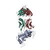
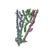
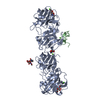
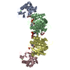
Mass: 23025.674 Da / Num. of mol.: 1 / Source method: isolated from a natural source / Details: MONOCLONAL ANTIBODY AGAINST HUMAN DDR1 / Source: (natural) MUS MUSCULUS (house mouse)
#3: Antibody
MONOCLONALANTIBODY3E3LIGHTCHAIN
Mass: 23410.799 Da / Num. of mol.: 1 / Source method: isolated from a natural source / Details: MONOCLONAL ANTIBODY AGAINST HUMAN DDR1 / Source: (natural) MUS MUSCULUS (house mouse)
-
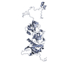
Protein / Sugars , 2 types, 3 molecules A
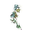
#1: Protein
EPITHELIALDISCOIDINDOMAIN-CONTAININGRECEPTOR1 / EPITHELIAL DISCOIDIN DOMAIN RECEPTOR 1 / CD167 ANTIGEN-LIKE FAMILY MEMBER A / CELL ADHESION KINASE ...EPITHELIAL DISCOIDIN DOMAIN RECEPTOR 1 / CD167 ANTIGEN-LIKE FAMILY MEMBER A / CELL ADHESION KINASE / DISCOIDIN RECEPTOR TYROSINE KINASE / HGK2 / MAMMARY CARCINOMA KINASE 10 / MCK-10 / PROTEIN-TYROSINE KINASE 3A / PROTEIN-TYROSINE KINASE RTK-6 / TRK E / TYROSINE KINASE DDR / TYROSINE-PROTEIN KINASE CAK / CD167A
Mass: 39354.312 Da / Num. of mol.: 1 / Fragment: RESIDUES 29-367 Source method: isolated from a genetically manipulated source Source: (gene. exp.) HOMO SAPIENS (human) / Plasmid: PCEP-PU / Cell line (production host): HEK293 C18 / Production host: HOMO SAPIENS (human) References: UniProt: Q08345, receptor protein-tyrosine kinase
In the structure databanks used in Yorodumi, some data are registered as the other names, "COVID-19 virus" and "2019-nCoV". Here are the details of the virus and the list of structure data.
Jan 31, 2019. EMDB accession codes are about to change! (news from PDBe EMDB page)
EMDB accession codes are about to change! (news from PDBe EMDB page)
The allocation of 4 digits for EMDB accession codes will soon come to an end. Whilst these codes will remain in use, new EMDB accession codes will include an additional digit and will expand incrementally as the available range of codes is exhausted. The current 4-digit format prefixed with “EMD-” (i.e. EMD-XXXX) will advance to a 5-digit format (i.e. EMD-XXXXX), and so on. It is currently estimated that the 4-digit codes will be depleted around Spring 2019, at which point the 5-digit format will come into force.
The EM Navigator/Yorodumi systems omit the EMD- prefix.
Related info.:Q: What is EMD? / ID/Accession-code notation in Yorodumi/EM Navigator
Yorodumi is a browser for structure data from EMDB, PDB, SASBDB, etc.
This page is also the successor to EM Navigator detail page, and also detail information page/front-end page for Omokage search.
The word "yorodu" (or yorozu) is an old Japanese word meaning "ten thousand". "mi" (miru) is to see.
Related info.:EMDB / PDB / SASBDB / Comparison of 3 databanks / Yorodumi Search / Aug 31, 2016. New EM Navigator & Yorodumi / Yorodumi Papers / Jmol/JSmol / Function and homology information / Changes in new EM Navigator and Yorodumi
 Movie
Movie Controller


 Open data
 Basic information
 Components
 Keywords
 Function and homology information
 HOMO SAPIENS (human)
 X-RAY DIFFRACTION /
  Authors
 Citation
 Structure visualization
 Downloads & links
 Other downloads
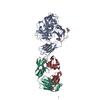

 PDBj
PDBj


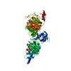
 Assembly
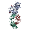


 Mass: 40.078 Da / Num. of mol.: 2 / Source method: obtained synthetically / Formula: Ca
 Sample preparation
 / Beamline: I02 / Wavelength: 0.9796
  Processing