 Movie
Movie Controller


+ Open data

- Basic information
| Entry | Database: PDB / ID: 6al5 | |||||||||
|---|---|---|---|---|---|---|---|---|---|---|
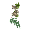
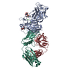
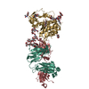
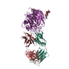
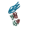
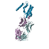
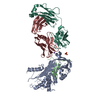
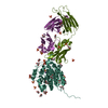
| Title | COMPLEX BETWEEN CD19 (N138Q MUTANT) AND B43 FAB | |||||||||
 Components Components |
| |||||||||
 Keywords Keywords | IMMUNE SYSTEM | |||||||||
| Function / homology |  Function and homology information Function and homology informationregulation of B cell activation / antigen receptor-mediated signaling pathway / B-1 B cell differentiation / regulation of B cell receptor signaling pathway / B cell proliferation involved in immune response / immunoglobulin mediated immune response / B cell proliferation / immunoglobulin complex / Regulation of Complement cascade / Antigen activates B Cell Receptor (BCR) leading to generation of second messengers ...regulation of B cell activation / antigen receptor-mediated signaling pathway / B-1 B cell differentiation / regulation of B cell receptor signaling pathway / B cell proliferation involved in immune response / immunoglobulin mediated immune response / B cell proliferation / immunoglobulin complex / Regulation of Complement cascade / Antigen activates B Cell Receptor (BCR) leading to generation of second messengers / positive regulation of release of sequestered calcium ion into cytosol / B cell receptor signaling pathway / Immunoregulatory interactions between a Lymphoid and a non-Lymphoid cell / Constitutive Signaling by Aberrant PI3K in Cancer / PIP3 activates AKT signaling / PI5P, PP2A and IER3 Regulate PI3K/AKT Signaling / adaptive immune response / positive regulation of phosphatidylinositol 3-kinase/protein kinase B signal transduction / membrane raft / external side of plasma membrane / protein-containing complex / extracellular exosome / extracellular region / plasma membrane Similarity search - Function | |||||||||
| Biological species |  Homo sapiens (human) Homo sapiens (human) | |||||||||
| Method |  X-RAY DIFFRACTION / SYNCHROTRON / MOLECULAR REPLACEMENT / Resolution: 3 Å X-RAY DIFFRACTION / SYNCHROTRON / MOLECULAR REPLACEMENT / Resolution: 3 Å | |||||||||
 Authors Authors | Teplyakov, A. / Obmolova, G. / Gilliland, G.L. | |||||||||
 Citation Citation | Journal: Proteins / Year: 2018 Title: Crystal structure of B-cell co-receptor CD19 in complex with antibody B43 reveals an unexpected fold. Authors: Teplyakov, A. / Obmolova, G. / Luo, J. / Gilliland, G.L. | |||||||||
| History |
|
- Structure visualization
| Structure viewer | Molecule: MolmilJmol/JSmol |
|---|
- Downloads & links
-Download
| PDBx/mmCIF format | 6al5.cif.gz | 144.1 KB | Display | PDBx/mmCIF format |
|---|---|---|---|---|
| PDB format | pdb6al5.ent.gz | 108.6 KB | Display | PDB format |
| PDBx/mmJSON format | 6al5.json.gz | Tree view | PDBx/mmJSON format | |
| Others |  Other downloads Other downloads |
-Validation report
| Arichive directory | https://data.pdbj.org/pub/pdb/validation_reports/al/6al5ftp://data.pdbj.org/pub/pdb/validation_reports/al/6al5 | HTTPS FTP |
|---|
-Related structure data
| Related structure data | 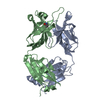 6al4C 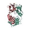 5i15S S: Starting model for refinement C: citing same article ( |
|---|---|
| Similar structure data |


-Links
 PDBj
PDBj


- Assembly
| Deposited unit | 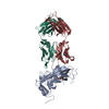
| ||||||||
|---|---|---|---|---|---|---|---|---|---|
| 1 |
| ||||||||
| Unit cell |
|
-Components
| #1: Protein | Mass: 29302.436 Da / Num. of mol.: 1 / Fragment: EXTRACELLULAR DOMAIN / Mutation: N138Q Source method: isolated from a genetically manipulated source Source: (gene. exp.) Homo sapiens (human) / Gene: CD19 / Production host:  unidentified baculovirus / References: UniProt: P15391 unidentified baculovirus / References: UniProt: P15391 | ||||
|---|---|---|---|---|---|
| #2: Antibody | Mass: 23815.260 Da / Num. of mol.: 1 Source method: isolated from a genetically manipulated source Source: (gene. exp.) Homo sapiens (human) / Gene: IGK@ / Production host: Homo sapiens (human) / References: UniProt: Q6P5S8 | ||||
| #3: Antibody | Mass: 25225.055 Da / Num. of mol.: 1 / Fragment: FD Source method: isolated from a genetically manipulated source Source: (gene. exp.) Homo sapiens (human) / Production host: Homo sapiens (human) / References: UniProt: A8K008 | ||||
| #4: Sugar |   Type: D-saccharide, beta linking / Mass: 221.208 Da / Num. of mol.: 2 Type: D-saccharide, beta linking / Mass: 221.208 Da / Num. of mol.: 2Source method: isolated from a genetically manipulated source Formula: C8H15NO6 #5: Water | ChemComp-HOH / |  Mass: 18.015 Da / Num. of mol.: 2 / Source method: isolated from a natural source / Formula: H2O Mass: 18.015 Da / Num. of mol.: 2 / Source method: isolated from a natural source / Formula: H2OHas protein modification | Y | |
-Experimental details
-Experiment
| Experiment | Method: X-RAY DIFFRACTION / Number of used crystals: 1 |
|---|
- Sample preparation
| Crystal | Density Matthews: 2.82 Å3/Da / Density % sol: 56 % |
|---|---|
| Crystal grow | Temperature: 293 K / Method: vapor diffusion, sitting drop / pH: 7.5 / Details: 0.1 M HEPES PH 7.5, 23% PEG 8000 / PH range: 7.5 |
-Data collection
| Diffraction | Mean temperature: 100 K |
|---|---|
| Diffraction source | Source: SYNCHROTRON / Site: APS  / Beamline: 17-ID / Wavelength: 1 Å / Beamline: 17-ID / Wavelength: 1 Å |
| Detector | Type: DECTRIS PILATUS 6M / Detector: PIXEL / Date: Oct 19, 2015 |
| Radiation | Protocol: SINGLE WAVELENGTH / Monochromatic (M) / Laue (L): M / Scattering type: x-ray |
| Radiation wavelength | Wavelength: 1 Å / Relative weight: 1 |
| Reflection | Resolution: 3→30 Å / Num. obs: 18262 / % possible obs: 99.5 % / Observed criterion σ(I): -3 / Redundancy: 6.4 % / Biso Wilson estimate: 59.6 Å2 / Rmerge(I) obs: 0.083 / Net I/σ(I): 18.4 |
| Reflection shell | Resolution: 3→3.08 Å / Redundancy: 5.8 % / Rmerge(I) obs: 0.496 / Mean I/σ(I) obs: 3.6 / Num. unique obs: 1285 / % possible all: 96.6 |
- Processing
| Software |
| ||||||||||||||||||||||||||||||||||||||||||||||||||||||||||||||||||||||||||||||||||||||||||||||||||||||||||||||||||||||||||||||||||||||||||||||||||||||||||||||||||||||||||||||||||||||
|---|---|---|---|---|---|---|---|---|---|---|---|---|---|---|---|---|---|---|---|---|---|---|---|---|---|---|---|---|---|---|---|---|---|---|---|---|---|---|---|---|---|---|---|---|---|---|---|---|---|---|---|---|---|---|---|---|---|---|---|---|---|---|---|---|---|---|---|---|---|---|---|---|---|---|---|---|---|---|---|---|---|---|---|---|---|---|---|---|---|---|---|---|---|---|---|---|---|---|---|---|---|---|---|---|---|---|---|---|---|---|---|---|---|---|---|---|---|---|---|---|---|---|---|---|---|---|---|---|---|---|---|---|---|---|---|---|---|---|---|---|---|---|---|---|---|---|---|---|---|---|---|---|---|---|---|---|---|---|---|---|---|---|---|---|---|---|---|---|---|---|---|---|---|---|---|---|---|---|---|---|---|---|---|
| Refinement | Method to determine structure: MOLECULAR REPLACEMENT Starting model: 5i15 Resolution: 3→20 Å / Cor.coef. Fo:Fc: 0.945 / Cor.coef. Fo:Fc free: 0.897 / SU B: 18.061 / SU ML: 0.327 / Cross valid method: THROUGHOUT / σ(F): 0 / ESU R Free: 0.436
| ||||||||||||||||||||||||||||||||||||||||||||||||||||||||||||||||||||||||||||||||||||||||||||||||||||||||||||||||||||||||||||||||||||||||||||||||||||||||||||||||||||||||||||||||||||||
| Solvent computation | Ion probe radii: 0.8 Å / Shrinkage radii: 0.8 Å / VDW probe radii: 1.2 Å | ||||||||||||||||||||||||||||||||||||||||||||||||||||||||||||||||||||||||||||||||||||||||||||||||||||||||||||||||||||||||||||||||||||||||||||||||||||||||||||||||||||||||||||||||||||||
| Displacement parameters | Biso mean: 63.5 Å2
| ||||||||||||||||||||||||||||||||||||||||||||||||||||||||||||||||||||||||||||||||||||||||||||||||||||||||||||||||||||||||||||||||||||||||||||||||||||||||||||||||||||||||||||||||||||||
| Refinement step | Cycle: LAST / Resolution: 3→20 Å
| ||||||||||||||||||||||||||||||||||||||||||||||||||||||||||||||||||||||||||||||||||||||||||||||||||||||||||||||||||||||||||||||||||||||||||||||||||||||||||||||||||||||||||||||||||||||
| Refine LS restraints |
|