| Entry | Database: PDB / ID: 4k2u
|
|---|
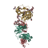
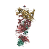
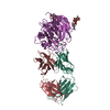
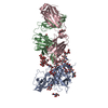
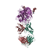
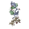
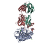
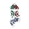
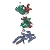
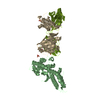
| Title | Crystal structure of PfEBA-175 F1 in complex with R218 antibody Fab fragment |
|---|
 Components Components | - Antibody Heavy Chain
- Antibody Light Chain
- Erythrocyte binding antigen 175
|
|---|
 Keywords Keywords | IMMUNE SYSTEM / DBL domain / Immunoglobulin domain / Antibody fragment / Cell attachment / Immunity / Receptor / Ligand / Extracellular |
|---|
| Function / homology |  Function and homology information Function and homology information
Erythrocyte binding antigen 175, C-terminal / Erythrocyte binding antigen 175, C-terminal domain superfamily / Erythrocyte binding antigen 175 / Duffy-antigen binding / Duffy-antigen binding superfamily / Duffy binding domain / Immunoglobulins / Immunoglobulin-like / Sandwich / Mainly BetaSimilarity search - Domain/homology |
|---|
| Biological species |   Plasmodium falciparum (malaria parasite P. falciparum) Plasmodium falciparum (malaria parasite P. falciparum)
  Mus musculus (house mouse) Mus musculus (house mouse) |
|---|
| Method |  X-RAY DIFFRACTION / SYNCHROTRON / MOLECULAR REPLACEMENT / Resolution: 2.45 Å X-RAY DIFFRACTION / SYNCHROTRON / MOLECULAR REPLACEMENT / Resolution: 2.45 Å |
|---|
 Authors Authors | Tolia, N.H. |
|---|
 Citation Citation | Journal: Plos Pathog. / Year: 2013
Title: Structural and Functional Basis for Inhibition of Erythrocyte Invasion by Antibodies that Target Plasmodium falciparum EBA-175.
Authors: Chen, E. / Paing, M.M. / Salinas, N. / Sim, B.K. / Tolia, N.H. |
|---|
| History | | Deposition | Apr 9, 2013 | Deposition site: RCSB / Processing site: RCSB |
|---|
| Revision 1.0 | Jun 12, 2013 | Provider: repository / Type: Initial release |
|---|
| Revision 1.1 | Nov 15, 2017 | Group: Refinement description / Category: software / Item: _software.name |
|---|
| Revision 1.2 | Nov 20, 2024 | Group: Data collection / Database references ...Data collection / Database references / Derived calculations / Structure summary
Category: chem_comp_atom / chem_comp_bond ...chem_comp_atom / chem_comp_bond / database_2 / pdbx_entry_details / pdbx_modification_feature / struct_site
Item: _database_2.pdbx_DOI / _database_2.pdbx_database_accession ..._database_2.pdbx_DOI / _database_2.pdbx_database_accession / _struct_site.pdbx_auth_asym_id / _struct_site.pdbx_auth_comp_id / _struct_site.pdbx_auth_seq_id |
|---|
|
|---|
 Movie
Movie Controller

 Yorodumi
 Open data
 Basic information
 Structure visualization
 Downloads & links
 Other downloads


 PDBj
PDBj

 Assembly
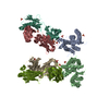


 Mass: 96.063 Da / Num. of mol.: 12 / Source method: obtained synthetically / Formula: SO4
 Mass: 18.015 Da / Num. of mol.: 171 / Source method: isolated from a natural source / Formula: H2O
 Sample preparation
 / Beamline: 19-ID / Wavelength: 0.9791212 Å
 Processing