 Movie
Movie Controller

[English] 日本語
 Yorodumi
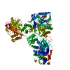
- PDB-4qex: Crystal structure of PfEBA-175 RII in complex with a Fab fragment... -
+ Open data

- Basic information
| Entry | Database: PDB / ID: 4qex | |||||||||
|---|---|---|---|---|---|---|---|---|---|---|
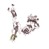
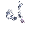
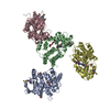
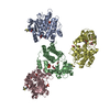
| Title | Crystal structure of PfEBA-175 RII in complex with a Fab fragment from inhibitory antibody R217 | |||||||||
 Components Components |
| |||||||||
 Keywords Keywords | IMMUNE SYSTEM / Duffy Binding Like (DBL) domain / Immunoglobulin domain / Invasion / adhesion / immunity / PFEBA-175 / Antibody / Cell Surface / Extrecellular / Cell Adhesion / Receptor / Ligand | |||||||||
| Function / homology | Erythrocyte binding antigen 175, C-terminal / Erythrocyte binding antigen 175, C-terminal domain superfamily / Erythrocyte binding antigen 175 / Duffy-antigen binding / Duffy-antigen binding superfamily / Duffy binding domain / host cell surface receptor binding / membrane / Erythrocyte-binding antigen-175 Function and homology information Function and homology information | |||||||||
| Biological species |   | |||||||||
| Method |  X-RAY DIFFRACTION / SYNCHROTRON / MOLECULAR REPLACEMENT / Resolution: 4.5 Å X-RAY DIFFRACTION / SYNCHROTRON / MOLECULAR REPLACEMENT / Resolution: 4.5 Å | |||||||||
 Authors Authors | Chen, E. / Paing, M.M. / Salinas, N. / Sim, B.K. / Tolia, N.H. | |||||||||
 Citation Citation | Journal: Plos Pathog. / Year: 2013 Title: Structural and Functional Basis for Inhibition of Erythrocyte Invasion by Antibodies that Target Plasmodium falciparum EBA-175. Authors: Chen, E. / Paing, M.M. / Salinas, N. / Sim, B.K. / Tolia, N.H. | |||||||||
| History |
|
- Structure visualization
| Structure viewer | Molecule: MolmilJmol/JSmol |
|---|
- Downloads & links
-Download
| PDBx/mmCIF format | 4qex.cif.gz | 668.2 KB | Display | PDBx/mmCIF format |
|---|---|---|---|---|
| PDB format | pdb4qex.ent.gz | 564.5 KB | Display | PDB format |
| PDBx/mmJSON format | 4qex.json.gz | Tree view | PDBx/mmJSON format | |
| Others |  Other downloads Other downloads |
-Validation report
| Arichive directory | https://data.pdbj.org/pub/pdb/validation_reports/qe/4qexftp://data.pdbj.org/pub/pdb/validation_reports/qe/4qex | HTTPS FTP |
|---|
-Related structure data


-Links
 PDBj
PDBj

- Assembly
| Deposited unit | 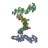
| ||||||||||||||||||||||||||||||||||||||||||||||
|---|---|---|---|---|---|---|---|---|---|---|---|---|---|---|---|---|---|---|---|---|---|---|---|---|---|---|---|---|---|---|---|---|---|---|---|---|---|---|---|---|---|---|---|---|---|---|---|
| 1 | 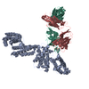
| ||||||||||||||||||||||||||||||||||||||||||||||
| 2 | 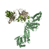
| ||||||||||||||||||||||||||||||||||||||||||||||
| Unit cell |
| ||||||||||||||||||||||||||||||||||||||||||||||
| Noncrystallographic symmetry (NCS) | NCS domain:
NCS domain segments:
NCS ensembles :
|
-Components
| #1: Protein | Mass: 72184.391 Da / Num. of mol.: 2 / Mutation: N3Q, S50A, S195A, T206A Source method: isolated from a genetically manipulated source Source: (gene. exp.) Gene: EBA-175 / Production host:  Komagataella pastoris (fungus) / References: UniProt: Q05644 Komagataella pastoris (fungus) / References: UniProt: Q05644#2: Antibody | Mass: 23622.992 Da / Num. of mol.: 2 / Source method: isolated from a natural source / Source: (natural) #3: Antibody | Mass: 22897.447 Da / Num. of mol.: 2 / Source method: isolated from a natural source / Source: (natural) Has protein modification | Y | |
|---|
-Experimental details
-Experiment
| Experiment | Method: X-RAY DIFFRACTION / Number of used crystals: 1 |
|---|
- Sample preparation
| Crystal | Density Matthews: 2.54 Å3/Da / Density % sol: 51.65 % |
|---|---|
| Crystal grow | Temperature: 290 K / Method: vapor diffusion, hanging drop / pH: 8 Details: 0.3 M magnesium chloride, 8% benzamidine hydrochloride, 7.5% glycerol, 18% PEG 3350, pH 8.0, VAPOR DIFFUSION, HANGING DROP, temperature 290K |
-Data collection
| Diffraction | Mean temperature: 93 K |
|---|---|
| Diffraction source | Source: SYNCHROTRON / Site: NSLS  / Beamline: X26C / Wavelength: 1.1 Å / Beamline: X26C / Wavelength: 1.1 Å |
| Detector | Type: ADSC QUANTUM 4 / Detector: CCD / Date: Apr 3, 2004 |
| Radiation | Protocol: SINGLE WAVELENGTH / Monochromatic (M) / Laue (L): M / Scattering type: x-ray |
| Radiation wavelength | Wavelength: 1.1 Å / Relative weight: 1 |
| Reflection | Resolution: 4.5→20 Å / Num. all: 14361 / Num. obs: 13322 / % possible obs: 92.8 % / Observed criterion σ(F): 0 / Observed criterion σ(I): 0 |
| Reflection shell | Resolution: 4.5→4.6 Å / % possible all: 52.8 |
- Processing
| Software |
| |||||||||||||||||||||||||||||||||||||||||||||||||
|---|---|---|---|---|---|---|---|---|---|---|---|---|---|---|---|---|---|---|---|---|---|---|---|---|---|---|---|---|---|---|---|---|---|---|---|---|---|---|---|---|---|---|---|---|---|---|---|---|---|---|
| Refinement | Method to determine structure: MOLECULAR REPLACEMENT / Resolution: 4.5→19.842 Å / SU ML: 0.63 / σ(F): 1.99 / Phase error: 31 / Stereochemistry target values: ML
| |||||||||||||||||||||||||||||||||||||||||||||||||
| Solvent computation | Shrinkage radii: 1.2 Å / VDW probe radii: 1.3 Å / Solvent model: FLAT BULK SOLVENT MODEL | |||||||||||||||||||||||||||||||||||||||||||||||||
| Refinement step | Cycle: LAST / Resolution: 4.5→19.842 Å
| |||||||||||||||||||||||||||||||||||||||||||||||||
| Refine LS restraints |
| |||||||||||||||||||||||||||||||||||||||||||||||||
| Refine LS restraints NCS |
| |||||||||||||||||||||||||||||||||||||||||||||||||
| LS refinement shell |
|
