 Movie
Movie Controller

[English] 日本語
 Yorodumi
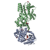
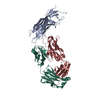
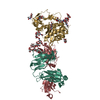
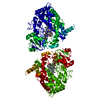
- PDB-6i3z: Fab fragment of an antibody selective for wild-type alpha-1-antit... -
+ Open data

- Basic information
| Entry | Database: PDB / ID: 6i3z | |||||||||||||||
|---|---|---|---|---|---|---|---|---|---|---|---|---|---|---|---|---|
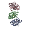
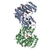
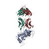
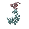
| Title | Fab fragment of an antibody selective for wild-type alpha-1-antitrypsin in complex with its antigen | |||||||||||||||
 Components Components |
| |||||||||||||||
 Keywords Keywords | PROTEIN BINDING / Antibody fragment / Antitrypsin binding / Diagnostic / Monoclonal / selective / wild-type / Glu342 / E342 | |||||||||||||||
| Function / homology |  Function and homology information Function and homology informationCargo concentration in the ER / COPII-coated ER to Golgi transport vesicle / COPII-mediated vesicle transport / endoplasmic reticulum-Golgi intermediate compartment membrane / platelet alpha granule lumen / acute-phase response / Post-translational protein phosphorylation / serine-type endopeptidase inhibitor activity / Regulation of Insulin-like Growth Factor (IGF) transport and uptake by Insulin-like Growth Factor Binding Proteins (IGFBPs) / blood coagulation ...Cargo concentration in the ER / COPII-coated ER to Golgi transport vesicle / COPII-mediated vesicle transport / endoplasmic reticulum-Golgi intermediate compartment membrane / platelet alpha granule lumen / acute-phase response / Post-translational protein phosphorylation / serine-type endopeptidase inhibitor activity / Regulation of Insulin-like Growth Factor (IGF) transport and uptake by Insulin-like Growth Factor Binding Proteins (IGFBPs) / blood coagulation / Platelet degranulation / extracellular matrix / protease binding / ficolin-1-rich granule lumen / endoplasmic reticulum lumen / Neutrophil degranulation / Golgi apparatus / endoplasmic reticulum / : / extracellular exosome / extracellular region / identical protein binding Similarity search - Function | |||||||||||||||
| Biological species |  Homo sapiens (human) Homo sapiens (human) | |||||||||||||||
| Method |  X-RAY DIFFRACTION / SYNCHROTRON / MOLECULAR REPLACEMENT / molecular replacement / Resolution: 3.1 Å X-RAY DIFFRACTION / SYNCHROTRON / MOLECULAR REPLACEMENT / molecular replacement / Resolution: 3.1 Å | |||||||||||||||
 Authors Authors | Laffranchi, M. / Elliston, E.L.K. / Miranda, E. / Perez, J. / Jagger, A.M. / Fra, A. / Lomas, D.A. / Irving, J.A. | |||||||||||||||
| Funding support |  United Kingdom, United Kingdom,  Italy, Italy,  United States, 4items United States, 4items
| |||||||||||||||
 Citation Citation | Journal: JCI Insight / Year: 2020 Title: Intrahepatic heteropolymerization of M and Z alpha-1-antitrypsin. Authors: Laffranchi, M. / Elliston, E.L. / Miranda, E. / Perez, J. / Ronzoni, R. / Jagger, A.M. / Heyer-Chauhan, N. / Brantly, M.L. / Fra, A. / Lomas, D.A. / Irving, J.A. | |||||||||||||||
| History |
|
- Structure visualization
| Structure viewer | Molecule: MolmilJmol/JSmol |
|---|
- Downloads & links
-Download
| PDBx/mmCIF format | 6i3z.cif.gz | 160.1 KB | Display | PDBx/mmCIF format |
|---|---|---|---|---|
| PDB format | pdb6i3z.ent.gz | 118.7 KB | Display | PDB format |
| PDBx/mmJSON format | 6i3z.json.gz | Tree view | PDBx/mmJSON format | |
| Others |  Other downloads Other downloads |
-Validation report
| Arichive directory | https://data.pdbj.org/pub/pdb/validation_reports/i3/6i3zftp://data.pdbj.org/pub/pdb/validation_reports/i3/6i3z | HTTPS FTP |
|---|
-Related structure data
| Related structure data |  6i1oC  1ezxS  6io1S S: Starting model for refinement C: citing same article ( |
|---|---|
| Similar structure data |


-Links
 PDBj
PDBj


- Assembly
| Deposited unit | 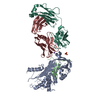
| ||||||||
|---|---|---|---|---|---|---|---|---|---|
| 1 |
| ||||||||
| Unit cell |
|
-Components
-Antibody , 2 types, 2 molecules HL
| #3: Antibody | Mass: 23129.936 Da / Num. of mol.: 1 / Source method: isolated from a natural source / Source: (natural) |
|---|---|
| #4: Antibody | Mass: 23914.398 Da / Num. of mol.: 1 / Source method: isolated from a natural source / Source: (natural) |
-Protein / Protein/peptide , 2 types, 2 molecules AB
| #1: Protein | Mass: 40018.207 Da / Num. of mol.: 1 Source method: isolated from a genetically manipulated source Source: (gene. exp.) Homo sapiens (human) / Gene: SERPINA1, AAT, PI, PRO0684, PRO2209 / Production host:  |
|---|---|
| #2: Protein/peptide | Mass: 4681.598 Da / Num. of mol.: 1 Source method: isolated from a genetically manipulated source Source: (gene. exp.) Homo sapiens (human) / Gene: SERPINA1, AAT, PI, PRO0684, PRO2209 / Production host: |
-Non-polymers , 3 types, 35 molecules 


| #5: Chemical |  Mass: 22.990 Da / Num. of mol.: 2 / Source method: obtained synthetically / Formula: Na Mass: 22.990 Da / Num. of mol.: 2 / Source method: obtained synthetically / Formula: Na#6: Chemical | ChemComp-SO4 / |  Mass: 96.063 Da / Num. of mol.: 1 / Source method: obtained synthetically / Formula: SO4 Mass: 96.063 Da / Num. of mol.: 1 / Source method: obtained synthetically / Formula: SO4#7: Water | ChemComp-HOH / | Mass: 18.015 Da / Num. of mol.: 32 / Source method: isolated from a natural source / Formula: H2O |
|---|
-Details
| Has protein modification | Y |
|---|
-Experimental details
-Experiment
| Experiment | Method: X-RAY DIFFRACTION / Number of used crystals: 1 |
|---|
- Sample preparation
| Crystal | Density Matthews: 2.37 Å3/Da / Density % sol: 48.12 % / Description: Plate |
|---|---|
| Crystal grow | Temperature: 289 K / Method: vapor diffusion, hanging drop / pH: 7.5 Details: 20% PEG 3350, 0.1 M ammonium sulfate, 0.1 M HEPES pH 7.5 |
-Data collection
| Diffraction | Mean temperature: 100 K / Ambient temp details: Cryostream / Serial crystal experiment: N | ||||||||||||||||||||||||
|---|---|---|---|---|---|---|---|---|---|---|---|---|---|---|---|---|---|---|---|---|---|---|---|---|---|
| Diffraction source | Source: SYNCHROTRON / Site: ESRF  / Beamline: ID29 / Wavelength: 1.0723 Å / Beamline: ID29 / Wavelength: 1.0723 Å | ||||||||||||||||||||||||
| Detector | Type: DECTRIS PILATUS3 6M / Detector: PIXEL / Date: Jul 12, 2018 / Details: Toroidal mirror | ||||||||||||||||||||||||
| Radiation | Monochromator: Si(111) / Protocol: SINGLE WAVELENGTH / Monochromatic (M) / Laue (L): M / Scattering type: x-ray | ||||||||||||||||||||||||
| Radiation wavelength | Wavelength: 1.0723 Å / Relative weight: 1 | ||||||||||||||||||||||||
| Reflection | Resolution: 3.1→37.82 Å / Num. obs: 13004 / % possible obs: 92.6 % / Redundancy: 3.3 % / Biso Wilson estimate: 51.51 Å2 / CC1/2: 0.965 / Rmerge(I) obs: 0.185 / Rpim(I) all: 0.127 / Rrim(I) all: 0.226 / Net I/σ(I): 3.4 | ||||||||||||||||||||||||
| Reflection shell | Diffraction-ID: 1
|
-Phasing
| Phasing | Method: molecular replacement | |||||||||
|---|---|---|---|---|---|---|---|---|---|---|
| Phasing MR |
|
- Processing
| Software |
| ||||||||||||||||||||||||||||||||||||
|---|---|---|---|---|---|---|---|---|---|---|---|---|---|---|---|---|---|---|---|---|---|---|---|---|---|---|---|---|---|---|---|---|---|---|---|---|---|
| Refinement | Method to determine structure: MOLECULAR REPLACEMENT Starting model: 6IO1,1EZX Resolution: 3.1→37.82 Å / SU ML: 0.52 / Cross valid method: THROUGHOUT / σ(F): 1.96 / Phase error: 30.66
| ||||||||||||||||||||||||||||||||||||
| Solvent computation | Shrinkage radii: 0.8 Å / VDW probe radii: 1 Å | ||||||||||||||||||||||||||||||||||||
| Displacement parameters | Biso max: 118.54 Å2 / Biso mean: 39.7541 Å2 / Biso min: 2.68 Å2 | ||||||||||||||||||||||||||||||||||||
| Refinement step | Cycle: final / Resolution: 3.1→37.82 Å
| ||||||||||||||||||||||||||||||||||||
| LS refinement shell | Refine-ID: X-RAY DIFFRACTION / Rfactor Rfree error: 0
|
