[English] 日本語
Yorodumi
- PDB-3v6o: Leptin Receptor-antibody complex -

+
Open data


ID or keywords:

Loading...

-
Basic information

Entry
Database: PDB / ID: 3v6o
TitleLeptin Receptor-antibody complex
Components
  • (Monoclonal antibody 9F8 fab fragment ...) x 2
  • Leptin receptor
Keywords
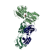
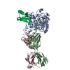
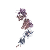
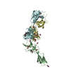
IMMUNE SYSTEM / Receptor-antibody complex / Cytokine receptor / Antibody Fab fragment / Immunoglobulin fold / Leptin Receptor-antibody complex
Function / homology
Function and homology information


leptin receptor activity / regulation of transport / bone growth / leptin-mediated signaling pathway / regulation of bone remodeling / response to leptin / regulation of feeding behavior / sexual reproduction / multicellular organism development / energy reserve metabolic process ...leptin receptor activity / regulation of transport / bone growth / leptin-mediated signaling pathway / regulation of bone remodeling / response to leptin / regulation of feeding behavior / sexual reproduction / multicellular organism development / energy reserve metabolic process / Signaling by Leptin / cytokine receptor activity / glycogen metabolic process / cytokine binding / transport across blood-brain barrier / T cell differentiation / glial cell proliferation / negative regulation of gluconeogenesis / phagocytosis / energy homeostasis / cholesterol metabolic process / negative regulation of autophagy / gluconeogenesis / cytokine-mediated signaling pathway / transmembrane signaling receptor activity / glucose homeostasis / positive regulation of cold-induced thermogenesis / basolateral plasma membrane / angiogenesis / cell surface receptor signaling pathway / receptor complex / positive regulation of protein phosphorylation / external side of plasma membrane / extracellular region / identical protein binding
Similarity search - Function
Leptin receptor, immunoglobulin-like domain / Obesity receptor immunoglobulin like domain / Immunoglobulin C2-set-like, ligand-binding / Ig-like C2-type domain / Short hematopoietin receptor, family 1, conserved site / Long hematopoietin receptor, Gp130 family 2, conserved site / Long hematopoietin receptor, gp130 family signature. / Fibronectin type 3 domain / Fibronectin type-III domain profile. / Fibronectin type III ...Leptin receptor, immunoglobulin-like domain / Obesity receptor immunoglobulin like domain / Immunoglobulin C2-set-like, ligand-binding / Ig-like C2-type domain / Short hematopoietin receptor, family 1, conserved site / Long hematopoietin receptor, Gp130 family 2, conserved site / Long hematopoietin receptor, gp130 family signature. / Fibronectin type 3 domain / Fibronectin type-III domain profile. / Fibronectin type III / Fibronectin type III superfamily / Ig-like domain profile. / Immunoglobulin-like domain / Immunoglobulins / Immunoglobulin-like fold / Immunoglobulin-like / Sandwich / Mainly Beta
Similarity search - Domain/homology
ACETATE ION / CYSTEINE / Leptin receptor
Similarity search - Component
Biological speciesHomo sapiens (human)
Mus musculus (house mouse)
MethodX-RAY DIFFRACTION / SYNCHROTRON / MOLECULAR REPLACEMENT / Resolution: 1.95 Å
AuthorsCarpenter, B. / Hemsworth, G.R. / Ross, R.J. / Artymiuk, P.J.
CitationJournal: Structure / Year: 2012
Title: Structure of the human obesity receptor leptin-binding domain reveals the mechanism of leptin antagonism by a monoclonal antibody.
Authors: Carpenter, B. / Hemsworth, G.R. / Wu, Z. / Maamra, M. / Strasburger, C.J. / Ross, R.J. / Artymiuk, P.J.
History
DepositionDec 20, 2011Deposition site: RCSB / Processing site: RCSB
Revision 1.0Mar 14, 2012Provider: repository / Type: Initial release
Revision 1.1Apr 25, 2012Group: Database references
Revision 1.2Jul 29, 2020Group: Data collection / Database references ...Data collection / Database references / Derived calculations / Structure summary
Category: chem_comp / entity ...chem_comp / entity / pdbx_chem_comp_identifier / pdbx_entity_nonpoly / pdbx_struct_conn_angle / struct_conn / struct_ref_seq / struct_site / struct_site_gen
Item: _chem_comp.name / _chem_comp.type ..._chem_comp.name / _chem_comp.type / _entity.pdbx_description / _pdbx_entity_nonpoly.name / _pdbx_struct_conn_angle.ptnr1_auth_seq_id / _pdbx_struct_conn_angle.ptnr3_auth_seq_id / _pdbx_struct_conn_angle.value / _struct_conn.conn_type_id / _struct_conn.id / _struct_conn.pdbx_dist_value / _struct_conn.pdbx_leaving_atom_flag / _struct_conn.pdbx_role / _struct_conn.ptnr1_auth_asym_id / _struct_conn.ptnr1_auth_comp_id / _struct_conn.ptnr1_auth_seq_id / _struct_conn.ptnr1_label_asym_id / _struct_conn.ptnr1_label_atom_id / _struct_conn.ptnr1_label_comp_id / _struct_conn.ptnr1_label_seq_id / _struct_conn.ptnr2_auth_asym_id / _struct_conn.ptnr2_auth_comp_id / _struct_conn.ptnr2_auth_seq_id / _struct_conn.ptnr2_label_asym_id / _struct_conn.ptnr2_label_atom_id / _struct_conn.ptnr2_label_comp_id / _struct_ref_seq.db_align_beg / _struct_ref_seq.db_align_end
Description: Carbohydrate remediation / Provider: repository / Type: Remediation
Revision 1.3Dec 27, 2023Group: Data collection / Database references ...Data collection / Database references / Derived calculations / Structure summary
Category: chem_comp / chem_comp_atom ...chem_comp / chem_comp_atom / chem_comp_bond / database_2 / struct_conn
Item: _chem_comp.pdbx_synonyms / _database_2.pdbx_DOI ..._chem_comp.pdbx_synonyms / _database_2.pdbx_DOI / _database_2.pdbx_database_accession / _struct_conn.conn_type_id / _struct_conn.id / _struct_conn.pdbx_dist_value / _struct_conn.pdbx_leaving_atom_flag / _struct_conn.ptnr1_auth_asym_id / _struct_conn.ptnr1_auth_seq_id / _struct_conn.ptnr1_label_asym_id / _struct_conn.ptnr1_label_seq_id / _struct_conn.ptnr2_auth_asym_id / _struct_conn.ptnr2_auth_seq_id / _struct_conn.ptnr2_label_asym_id / _struct_conn.ptnr2_label_seq_id

-
Structure visualization

Structure viewerMolecule:
MolmilJmol/JSmol

Downloads & links

-
Assembly

Deposited unit
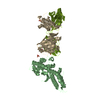
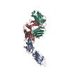
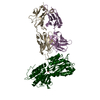
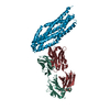
A: Leptin receptor
B: Leptin receptor
C: Monoclonal antibody 9F8 fab fragment Heavy chain
D: Monoclonal antibody 9F8 fab fragment Heavy chain
E: Monoclonal antibody 9F8 fab fragment Light chain
F: Monoclonal antibody 9F8 fab fragment Light chain
hetero molecules


Theoretical massNumber of molelcules
Total (without water)144,45647
Polymers141,6916
Non-polymers2,76541
Water15,331851
1
A: Leptin receptor
C: Monoclonal antibody 9F8 fab fragment Heavy chain
E: Monoclonal antibody 9F8 fab fragment Light chain
hetero molecules


Theoretical massNumber of molelcules
Total (without water)71,97421
Polymers70,8463
Non-polymers1,12818
Water543
TypeNameSymmetry operationNumber
identity operation1_555x,y,z1
Buried area5220 Å2
ΔGint-33 kcal/mol
Surface area29270 Å2
MethodPISA
2
B: Leptin receptor
D: Monoclonal antibody 9F8 fab fragment Heavy chain
F: Monoclonal antibody 9F8 fab fragment Light chain
hetero molecules


Theoretical massNumber of molelcules
Total (without water)72,48226
Polymers70,8463
Non-polymers1,63723
Water543
TypeNameSymmetry operationNumber
identity operation1_555x,y,z1
Buried area5350 Å2
ΔGint-31 kcal/mol
Surface area29250 Å2
MethodPISA
Unit cell
Length a, b, c (Å)89.814, 118.829, 171.281
Angle α, β, γ (deg.)90.000, 90.000, 90.000
Int Tables number19
Space group name H-MP212121

-
Components

-
Antibody , 2 types, 4 molecules CDEF

#2: Antibody Monoclonal antibody 9F8 fab fragment Heavy chain


Mass: 23592.359 Da / Num. of mol.: 2 / Fragment: Fab fragment of monoclonal antibody H chain / Source method: isolated from a natural source / Source: (natural) Mus musculus (house mouse)
#3: Antibody Monoclonal antibody 9F8 fab fragment Light chain


Mass: 23873.506 Da / Num. of mol.: 2 / Fragment: Fab fragment of monoclonal antibody L chain / Source method: isolated from a natural source / Source: (natural) Mus musculus (house mouse)

-
Protein / Sugars , 2 types, 3 molecules AB

#1: Protein Leptin receptor / / LEP-R / HuB219 / OB receptor / OB-R


Mass: 23379.795 Da / Num. of mol.: 2 / Fragment: Leptin binding domain of human obesity receptor
Source method: isolated from a genetically manipulated source
Source: (gene. exp.) Homo sapiens (human) / Gene: DB, LEPR, OBR / Plasmid: pET21a(+) / Production host: Escherichia coli (E. coli) / Strain (production host): BL21(DE3)-RIPL / References: UniProt: P48357
#8: Sugar ChemComp-NAG / 2-acetamido-2-deoxy-beta-D-glucopyranose / N-acetyl-beta-D-glucosamine / 2-acetamido-2-deoxy-beta-D-glucose / 2-acetamido-2-deoxy-D-glucose / 2-acetamido-2-deoxy-glucose / N-ACETYL-D-GLUCOSAMINE / N-Acetylglucosamine


Type: D-saccharide, beta linking / Mass: 221.208 Da / Num. of mol.: 1
Source method: isolated from a genetically manipulated source
Formula: C8H15NO6
IdentifierTypeProgram
DGlcpNAcbCONDENSED IUPAC CARBOHYDRATE SYMBOLGMML 1.0
N-acetyl-b-D-glucopyranosamineCOMMON NAMEGMML 1.0
b-D-GlcpNAcIUPAC CARBOHYDRATE SYMBOLPDB-CARE 1.0
GlcNAcSNFG CARBOHYDRATE SYMBOLGMML 1.0

-
Non-polymers , 5 types, 891 molecules

#4: Chemical ChemComp-CYS / CYSTEINE / Cysteine


Type: L-peptide linking / Mass: 121.158 Da / Num. of mol.: 2 / Source method: obtained synthetically / Formula: C3H7NO2S
#5: Chemical...
ChemComp-EDO / 1,2-ETHANEDIOL / ETHYLENE GLYCOL / Ethylene glycol


Mass: 62.068 Da / Num. of mol.: 31 / Source method: obtained synthetically / Formula: C2H6O2
#6: Chemical
ChemComp-ACT / ACETATE ION / Acetate


Mass: 59.044 Da / Num. of mol.: 6 / Source method: obtained synthetically / Formula: C2H3O2
#7: Chemical ChemComp-NA / SODIUM ION


Mass: 22.990 Da / Num. of mol.: 1 / Source method: obtained synthetically / Formula: Na
#9: Water ChemComp-HOH / water / Water


Mass: 18.015 Da / Num. of mol.: 851 / Source method: isolated from a natural source / Formula: H2O

-
Experimental details

-
Experiment

ExperimentMethod: X-RAY DIFFRACTION / Number of used crystals: 1

-
Sample preparation

CrystalDensity Matthews: 3.22 Å3/Da / Density % sol: 61.81 %
Crystal growTemperature: 281 K / Method: vapor diffusion / pH: 4.6
Details: sodium acetate, PEG-4000, pH 4.6, vapor diffusion, temperature 281K

-
Data collection

DiffractionMean temperature: 100 K
Diffraction sourceSource: SYNCHROTRON / Site: Diamond / Beamline: I02 / Wavelength: 0.9796 Å
DetectorType: ADSC QUANTUM 315 / Detector: CCD / Date: Apr 5, 2009
RadiationProtocol: SINGLE WAVELENGTH / Monochromatic (M) / Laue (L): M / Scattering type: x-ray
Radiation wavelengthWavelength: 0.9796 Å / Relative weight: 1
ReflectionResolution: 1.95→49 Å / Num. all: 127050 / Num. obs: 127050 / % possible obs: 99.9 % / Observed criterion σ(F): 0 / Observed criterion σ(I): 0 / Biso Wilson estimate: 22.1 Å2 / Rmerge(I) obs: 0.074 / Rsym value: 0.074 / Net I/σ(I): 15.9
Reflection shellResolution: 1.95→2 Å / Rmerge(I) obs: 0.556 / Mean I/σ(I) obs: 3.8 / % possible all: 99.9

-
Processing

Software
NameVersionClassificationNB
REFMAC5.5.0109refinement
PDB_EXTRACT3.1data extraction
ADSCQuantumdata collection
MOSFLMdata reduction
SCALAdata scaling
PHASERphasing
RefinementMethod to determine structure: MOLECULAR REPLACEMENT / Resolution: 1.95→49 Å / Cor.coef. Fo:Fc: 0.964 / Cor.coef. Fo:Fc free: 0.947 / Occupancy max: 1 / Occupancy min: 1 / SU B: 6.891 / SU ML: 0.088 / Cross valid method: THROUGHOUT / σ(F): 0 / ESU R: 0.12 / ESU R Free: 0.122 / Stereochemistry target values: MAXIMUM LIKELIHOOD
Details: HYDROGENS HAVE BEEN ADDED IN THE RIDING POSITIONS; U VALUES: WITH TLS ADDED
RfactorNum. reflection% reflectionSelection details
Rfree0.2135 6729 5 %RANDOM
Rwork0.1714 ---
all0.1735 127050 --
obs0.1735 -99.92 %-
Solvent computationIon probe radii: 0.8 Å / Shrinkage radii: 0.8 Å / VDW probe radii: 1.4 Å / Solvent model: MASK
Displacement parametersBiso max: 117.01 Å2 / Biso mean: 38.3931 Å2 / Biso min: 12.06 Å2
Baniso -1Baniso -2Baniso -3
1-3.06 Å20 Å20 Å2
2---0.58 Å20 Å2
3----2.48 Å2
Refinement stepCycle: LAST / Resolution: 1.95→49 Å
ProteinNucleic acidLigandSolventTotal
Num. atoms9486 0 177 851 10514
Refine LS restraints
Refine-IDTypeDev idealDev ideal targetNumber
X-RAY DIFFRACTIONr_bond_refined_d0.0230.0229879
X-RAY DIFFRACTIONr_bond_other_d0.0020.026643
X-RAY DIFFRACTIONr_angle_refined_deg1.841.95913399
X-RAY DIFFRACTIONr_angle_other_deg0.971316236
X-RAY DIFFRACTIONr_dihedral_angle_1_deg7.59351209
X-RAY DIFFRACTIONr_dihedral_angle_2_deg35.73324.256383
X-RAY DIFFRACTIONr_dihedral_angle_3_deg13.547151572
X-RAY DIFFRACTIONr_dihedral_angle_4_deg19.7481538
X-RAY DIFFRACTIONr_chiral_restr0.1260.21509
X-RAY DIFFRACTIONr_gen_planes_refined0.010.02110720
X-RAY DIFFRACTIONr_gen_planes_other0.0010.021893
X-RAY DIFFRACTIONr_mcbond_it3.761.56134
X-RAY DIFFRACTIONr_mcbond_other1.5351.52449
X-RAY DIFFRACTIONr_mcangle_it5.19829982
X-RAY DIFFRACTIONr_scbond_it7.3433745
X-RAY DIFFRACTIONr_scangle_it9.2974.53417
LS refinement shellResolution: 1.95→2.001 Å / Total num. of bins used: 20
RfactorNum. reflection% reflection
Rfree0.289 494 -
Rwork0.254 9261 -
all-9755 -
obs--99.94 %
Refinement TLS params.

Method: refined / Refine-ID: X-RAY DIFFRACTION

IDL112)L122)L132)L222)L232)L332)S11 (Å °)S12 (Å °)S13 (Å °)S21 (Å °)S22 (Å °)S23 (Å °)S31 (Å °)S32 (Å °)S33 (Å °)T112)T122)T132)T222)T232)T332)Origin x (Å)Origin y (Å)Origin z (Å)
10.5756-0.3849-0.05490.27650.16991.3628-0.0086-0.01270.01530.04720.0446-0.00060.3160.1176-0.0360.16440.026-0.00760.09280.00210.049847.140140.3999-6.3344
26.2909-0.77613.69010.104-0.45232.1677-0.16610.20460.20640.02940.0249-0.0354-0.11070.13430.14130.05350.0272-0.07120.1912-0.03670.094871.68849.845216.4967
30.30860.5091-0.15720.8811-0.09841.33730.02770.01580.01480.00940.01420.0545-0.3762-0.06-0.04180.20510.03960.01630.0489-0.00110.058540.494869.7295.7121
47.85275.0629-5.6473.2828-3.58664.2211-0.25080.17-0.3972-0.22020.0877-0.293-0.0185-0.15630.1630.3189-0.05790.14470.10630.00850.093466.644570.8737-17.5072
50.19040.0514-0.00070.4135-0.14131.93380.0062-0.0466-0.0247-0.09320.01250.01590.00220.067-0.01880.05360.0031-0.00790.09760.010.061642.184351.5662-31.9768
60.10570.4594-0.00992.0165-0.09640.3970.0039-0.0165-0.01560.0468-0.0975-0.10450.03070.07430.09360.0831-0.0033-0.03030.0680.02790.097544.087779.6554-47.4473
70.12330.293-0.04430.7446-0.24351.47360.03970.0335-0.00950.16820.0382-0.0007-0.1566-0.0604-0.07790.14690.0120.01840.0744-0.00280.04941.738856.309531.0013
80.1016-0.4434-0.24592.63760.29061.62230.0320.00330.0001-0.0564-0.2219-0.256-0.24610.20750.18980.078-0.0549-0.01050.07790.07360.146253.754631.674245.9256
90.0397-0.12380.1591.2691-0.31010.9642-0.0404-0.0395-0.03130.08660.08350.0082-0.2556-0.2655-0.04310.08770.05770.00550.14460.02040.073130.699864.2918-18.272
100.2365-0.1097-0.22140.6937-0.70471.6005-0.18060.02210.0580.13030.1236-0.0116-0.0069-0.10270.0570.1881-0.0205-0.0660.06150.0510.074730.280386.3012-48.7786
110.0442-0.26980.06141.7518-0.59811.00090.0021-0.01150.004-0.09380.07380.03410.1854-0.176-0.07590.0742-0.0255-0.00130.09940.01140.067436.00840.192217.3735
120.09530.09870.30131.0711-0.61281.8592-0.0302-0.017-0.0138-0.0308-0.0008-0.0638-0.0408-0.0540.0310.13450.01860.01240.04080.04040.073443.524319.591147.5619
Refinement TLS group
IDRefine-IDRefine TLS-IDAuth asym-IDAuth seq-ID
1X-RAY DIFFRACTION1A431 - 533
2X-RAY DIFFRACTION2A534 - 701
3X-RAY DIFFRACTION3B430 - 533
4X-RAY DIFFRACTION4B534 - 1001
5X-RAY DIFFRACTION5C3 - 119
6X-RAY DIFFRACTION6C120 - 222
7X-RAY DIFFRACTION7D3 - 119
8X-RAY DIFFRACTION8D120 - 221
9X-RAY DIFFRACTION9E4 - 106
10X-RAY DIFFRACTION10E107 - 216
11X-RAY DIFFRACTION11F4 - 106
12X-RAY DIFFRACTION12F107 - 216

+
About Yorodumi

-
News

-
Feb 9, 2022. New format data for meta-information of EMDB entries

New format data for meta-information of EMDB entries

  • Version 3 of the EMDB header file is now the official format.
  • The previous official version 1.9 will be removed from the archive.

Related info.:EMDB header

External links:wwPDB to switch to version 3 of the EMDB data model

-
Aug 12, 2020. Covid-19 info

Covid-19 info

URL: https://pdbj.org/emnavi/covid19.php

New page: Covid-19 featured information page in EM Navigator.

Related info.:Covid-19 info / Mar 5, 2020. Novel coronavirus structure data

+
Mar 5, 2020. Novel coronavirus structure data

Novel coronavirus structure data

Related info.:Yorodumi Speices / Aug 12, 2020. Covid-19 info

External links:COVID-19 featured content - PDBj / Molecule of the Month (242):Coronavirus Proteases

+
Jan 31, 2019. EMDB accession codes are about to change! (news from PDBe EMDB page)

EMDB accession codes are about to change! (news from PDBe EMDB page)

  • The allocation of 4 digits for EMDB accession codes will soon come to an end. Whilst these codes will remain in use, new EMDB accession codes will include an additional digit and will expand incrementally as the available range of codes is exhausted. The current 4-digit format prefixed with “EMD-” (i.e. EMD-XXXX) will advance to a 5-digit format (i.e. EMD-XXXXX), and so on. It is currently estimated that the 4-digit codes will be depleted around Spring 2019, at which point the 5-digit format will come into force.
  • The EM Navigator/Yorodumi systems omit the EMD- prefix.

Related info.:Q: What is EMD? / ID/Accession-code notation in Yorodumi/EM Navigator

External links:EMDB Accession Codes are Changing Soon! / Contact to PDBj

+
Jul 12, 2017. Major update of PDB

Major update of PDB

  • wwPDB released updated PDB data conforming to the new PDBx/mmCIF dictionary.
  • This is a major update changing the version number from 4 to 5, and with Remediation, in which all the entries are updated.
  • In this update, many items about electron microscopy experimental information are reorganized (e.g. em_software).
  • Now, EM Navigator and Yorodumi are based on the updated data.

External links:wwPDB Remediation / Enriched Model Files Conforming to OneDep Data Standards Now Available in the PDB FTP Archive

-
Yorodumi

Thousand views of thousand structures

  • Yorodumi is a browser for structure data from EMDB, PDB, SASBDB, etc.
  • This page is also the successor to EM Navigator detail page, and also detail information page/front-end page for Omokage search.
  • The word "yorodu" (or yorozu) is an old Japanese word meaning "ten thousand". "mi" (miru) is to see.

Related info.:EMDB / PDB / SASBDB / Comparison of 3 databanks / Yorodumi Search / Aug 31, 2016. New EM Navigator & Yorodumi / Yorodumi Papers / Jmol/JSmol / Function and homology information / Changes in new EM Navigator and Yorodumi

Read more