 Movie
Movie Controller

[English] 日本語
 Yorodumi
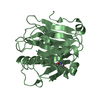
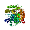
- PDB-1ehy: X-ray structure of the epoxide hydrolase from agrobacterium radio... -
+ Open data

- Basic information
| Entry | Database: PDB / ID: 1ehy | ||||||
|---|---|---|---|---|---|---|---|
| Title | X-ray structure of the epoxide hydrolase from agrobacterium radiobacter ad1 | ||||||
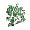
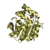
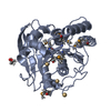
 Components Components | PROTEIN (SOLUBLE EPOXIDE HYDROLASE) | ||||||
 Keywords Keywords | HYDROLASE / ALPHA/BETA HYDROLASE FOLD / EPOXIDE DEGRADATION / EPICHLOROHYDRIN | ||||||
| Function / homology |  Function and homology information Function and homology information | ||||||
| Biological species |  Agrobacterium tumefaciens (bacteria) Agrobacterium tumefaciens (bacteria) | ||||||
| Method |  X-RAY DIFFRACTION / SYNCHROTRON / SIRAS / Resolution: 2.1 Å X-RAY DIFFRACTION / SYNCHROTRON / SIRAS / Resolution: 2.1 Å | ||||||
 Authors Authors | Nardini, M. / Ridder, I.S. / Rozeboom, H.J. / Kalk, K.H. / Rink, R. / Janssen, D.B. / Dijkstra, B.W. | ||||||
 Citation Citation | Journal: J.Biol.Chem. / Year: 1999 Title: The x-ray structure of epoxide hydrolase from Agrobacterium radiobacter AD1. An enzyme to detoxify harmful epoxides. Authors: Nardini, M. / Ridder, I.S. / Rozeboom, H.J. / Kalk, K.H. / Rink, R. / Janssen, D.B. / Dijkstra, B.W. #1: Journal: Tetrahedron / Year: 1998Title: Enantioselectivity of a Recombinant Epoxide Hydrolase from Agrobacterium Radiobacter Authors: Lutje Spelberg, J.H. / Rink, R. / Kellogg, R.M. / Janssen, D.B. #2: Journal: J.Biol.Chem. / Year: 1997Title: Primary Structure and Catalytic Mechanism of the Epoxide Hydrolase from Agrobacterium Radiobacter AD1 Authors: Rink, R. / Fennema, M. / Smids, M. / Dehmel, U. / Janssen, D.B. #3: Journal: Biochemistry / Year: 1998Title: Kinetic Mechanism of the Enantioselective Conversion of Styrene Oxide by Epoxide Hydrolase from Agrobacterium Radiodacter AD1 Authors: Jacobs, M.H.J. / Van Den Wijngaard, A.J. / Pentenga, M. / Janssen, D.B. #4: Journal: To be PublishedTitle: Mutation of Tyrosine Residues Involved in the Alkylation Half Reaction of Epoxide Hydrolase from Agrobacterium Radiodacter AD1 Results in Improved Enantioselectivity Authors: Rink, R. / Lutje Spelberg, J.H. / Pieters, R.J. / Kingma, J. / Nardini, M. / Kellogg, R.M. / Dijkstra, B.W. / Janssen, D.B. | ||||||
| History |
|
- Structure visualization
| Structure viewer | Molecule: MolmilJmol/JSmol |
|---|
- Downloads & links
-Download
| PDBx/mmCIF format | 1ehy.cif.gz | 248 KB | Display | PDBx/mmCIF format |
|---|---|---|---|---|
| PDB format | pdb1ehy.ent.gz | 201.2 KB | Display | PDB format |
| PDBx/mmJSON format | 1ehy.json.gz | Tree view | PDBx/mmJSON format | |
| Others |  Other downloads Other downloads |
-Validation report
| Arichive directory | https://data.pdbj.org/pub/pdb/validation_reports/eh/1ehyftp://data.pdbj.org/pub/pdb/validation_reports/eh/1ehy | HTTPS FTP |
|---|
-Related structure data
| Similar structure data |
|---|


-Links
 PDBj
PDBj


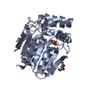
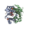
- Assembly
| Deposited unit | 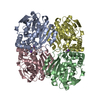
| ||||||||||||||||
|---|---|---|---|---|---|---|---|---|---|---|---|---|---|---|---|---|---|
| 1 | 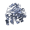
| ||||||||||||||||
| 2 | 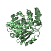
| ||||||||||||||||
| 3 | 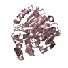
| ||||||||||||||||
| 4 | 
| ||||||||||||||||
| Unit cell |
| ||||||||||||||||
| Noncrystallographic symmetry (NCS) | NCS oper:
|
-Components
| #1: Protein | Mass: 34074.656 Da / Num. of mol.: 4 Source method: isolated from a genetically manipulated source Source: (gene. exp.) Agrobacterium tumefaciens (bacteria) / Strain: AD1 / Plasmid: PGELAF+ / Species (production host): Escherichia coli / Production host: #2: Chemical | ChemComp-K /   Mass: 39.098 Da / Num. of mol.: 4 / Source method: obtained synthetically / Formula: K Mass: 39.098 Da / Num. of mol.: 4 / Source method: obtained synthetically / Formula: K#3: Water | ChemComp-HOH / |  Mass: 18.015 Da / Num. of mol.: 577 / Source method: isolated from a natural source / Formula: H2O Mass: 18.015 Da / Num. of mol.: 577 / Source method: isolated from a natural source / Formula: H2OCompound details | DUE TO A CRYSTAL CONTACT BETWEEN LOOP RESIDUES 246-251 OF MONOMERS C AND D AND MONOMERS A AND B, IN ...DUE TO A CRYSTAL CONTACT BETWEEN LOOP RESIDUES 246-251 OF MONOMERS C AND D AND MONOMERS A AND B, IN ALL 4 MONOMERS THE CATALYTIC ASP 246 IS PULLED OUT FROM THE ACTIVE SITE, SO ALLOWING RESIDUE GLN 134 TO MOVE INTO THE ACTIVE SITE. | Has protein modification | Y | |
|---|
-Experimental details
-Experiment
| Experiment | Method: X-RAY DIFFRACTION / Number of used crystals: 1 |
|---|
- Sample preparation
| Crystal | Density Matthews: 2.57 Å3/Da / Density % sol: 52 % | |||||||||||||||
|---|---|---|---|---|---|---|---|---|---|---|---|---|---|---|---|---|
| Crystal grow | pH: 7 / Details: 1.8 M KH2PO4/K2HPO4, pH 7.0 | |||||||||||||||
| Crystal grow | *PLUS Method: vapor diffusion, hanging drop | |||||||||||||||
| Components of the solutions | *PLUS
|
-Data collection
| Diffraction | Mean temperature: 100 K |
|---|---|
| Diffraction source | Source: SYNCHROTRON / Site: ELETTRA  / Beamline: 5.2R / Wavelength: 1 / Beamline: 5.2R / Wavelength: 1 |
| Detector | Type: MARRESEARCH / Detector: IMAGE PLATE / Date: Aug 15, 1997 |
| Radiation | Protocol: SINGLE WAVELENGTH / Monochromatic (M) / Laue (L): M / Scattering type: x-ray |
| Radiation wavelength | Wavelength: 1 Å / Relative weight: 1 |
| Reflection | Resolution: 2.1→25 Å / Num. obs: 73445 / % possible obs: 91.5 % / Observed criterion σ(I): 0 / Redundancy: 3 % / Rmerge(I) obs: 0.062 / Net I/σ(I): 14.7 |
| Reflection shell | Resolution: 2.1→2.14 Å / Rmerge(I) obs: 0.36 / Mean I/σ(I) obs: 2.57 / % possible all: 82 |
| Reflection | *PLUS Num. measured all: 222880 |
| Reflection shell | *PLUS % possible obs: 82 % |
- Processing
| Software |
| ||||||||||||||||||||
|---|---|---|---|---|---|---|---|---|---|---|---|---|---|---|---|---|---|---|---|---|---|
| Refinement | Method to determine structure: SIRAS / Resolution: 2.1→25 Å / Rfactor Rfree error: 0.004 / Data cutoff high absF: 1000000 / Data cutoff low absF: 0.001 / Cross valid method: THROUGHOUT / σ(F): 0 Details: BULK SOLVENT CORRECTION USED DURING THE REFINEMENT IN ALL 4 MOLECULES IN THE ASYMMETRIC UNIT THE LOOP 246-251 SHOWS POOR ELECTRON DENSITY. HOWEVER CLEAR ELECTRON DENSITY IS PRESENT FOR THE ...Details: BULK SOLVENT CORRECTION USED DURING THE REFINEMENT IN ALL 4 MOLECULES IN THE ASYMMETRIC UNIT THE LOOP 246-251 SHOWS POOR ELECTRON DENSITY. HOWEVER CLEAR ELECTRON DENSITY IS PRESENT FOR THE SS BOND BETWEEN RESIDUES C248 AND D248. DUE TO A SLIGHTLY DIFFERENT CONFORMATION OF THAT LOOP IN MONOMER A, NO SS BOND IS OBSERVED BETWEEN RESIDUES A248 AND B248
| ||||||||||||||||||||
| Displacement parameters | Biso mean: 28.3 Å2 | ||||||||||||||||||||
| Refine analyze |
| ||||||||||||||||||||
| Refinement step | Cycle: LAST / Resolution: 2.1→25 Å
| ||||||||||||||||||||
| Refine LS restraints |
| ||||||||||||||||||||
| Refine LS restraints NCS | NCS model details: RESTRAINTS | ||||||||||||||||||||
| LS refinement shell | Resolution: 2.1→2.16 Å / Rfactor Rfree error: 0.022 / Total num. of bins used: 12
| ||||||||||||||||||||
| Xplor file |
| ||||||||||||||||||||
| Software | *PLUS Name: X-PLOR / Version: 3.843 / Classification: refinement | ||||||||||||||||||||
| Refinement | *PLUS Highest resolution: 2.1 Å / Lowest resolution: 25 Å / σ(F): 0 / % reflection Rfree: 5.2 % / Rfactor obs: 0.19 / Rfactor Rwork: 0.19 | ||||||||||||||||||||
| Solvent computation | *PLUS | ||||||||||||||||||||
| Displacement parameters | *PLUS Biso mean: 28.3 Å2 | ||||||||||||||||||||
| Refine LS restraints | *PLUS
| ||||||||||||||||||||
| LS refinement shell | *PLUS Highest resolution: 2.1 Å / Rfactor Rfree: 0.318 / % reflection Rfree: 4.5 % / Rfactor Rwork: 0.286 |
