 Movie
Movie Controller

[English] 日本語
 Yorodumi
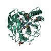
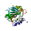
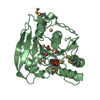
- PDB-6eic: Crystal structure of Rv0183, a Monoglyceride Lipase from Mycobact... -
+ Open data

- Basic information
| Entry | Database: PDB / ID: 6eic | ||||||
|---|---|---|---|---|---|---|---|
| Title | Crystal structure of Rv0183, a Monoglyceride Lipase from Mycobacterium Tuberculosis | ||||||
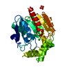
 Components Components | Mycobacterium Tuberculosis Monoglyceride Lipase | ||||||
 Keywords Keywords | HYDROLASE / Monoglycerid Lipase / Lipase / Alpha/Beta hydrolase | ||||||
| Function / homology |  Function and homology information Function and homology informationglycerolipid catabolic process / acylglycerol lipase / lipase activity / monoacylglycerol lipase activity / peptidoglycan-based cell wall / symbiont-mediated activation of host apoptosis / extracellular region / membrane / plasma membrane Similarity search - Function | ||||||
| Biological species |  Mycobacterium tuberculosis H37Rv (bacteria) Mycobacterium tuberculosis H37Rv (bacteria) | ||||||
| Method |  X-RAY DIFFRACTION / SYNCHROTRON / MOLECULAR REPLACEMENT / Resolution: 1.8 Å X-RAY DIFFRACTION / SYNCHROTRON / MOLECULAR REPLACEMENT / Resolution: 1.8 Å | ||||||
 Authors Authors | Aschauer, P. / Pavkov-Keller, T. / Oberer, M. | ||||||
| Funding support |  Austria, 1items Austria, 1items
| ||||||
 Citation Citation | Journal: Sci Rep / Year: 2018 Title: The crystal structure of monoacylglycerol lipase from M. tuberculosis reveals the basis for specific inhibition. Authors: Aschauer, P. / Zimmermann, R. / Breinbauer, R. / Pavkov-Keller, T. / Oberer, M. | ||||||
| History |
|
- Structure visualization
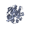
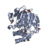
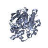
| Structure viewer | Molecule: MolmilJmol/JSmol |
|---|
- Downloads & links
-Download
| PDBx/mmCIF format | 6eic.cif.gz | 174.2 KB | Display | PDBx/mmCIF format |
|---|---|---|---|---|
| PDB format | pdb6eic.ent.gz | 139.6 KB | Display | PDB format |
| PDBx/mmJSON format | 6eic.json.gz | Tree view | PDBx/mmJSON format | |
| Others |  Other downloads Other downloads |
-Validation report
| Arichive directory | https://data.pdbj.org/pub/pdb/validation_reports/ei/6eicftp://data.pdbj.org/pub/pdb/validation_reports/ei/6eic | HTTPS FTP |
|---|
-Related structure data
| Similar structure data |
|---|


-Links
 PDBj
PDBj

- Assembly
| Deposited unit | 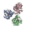
| ||||||||
|---|---|---|---|---|---|---|---|---|---|
| 1 | 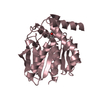
| ||||||||
| 2 | 
| ||||||||
| 3 | 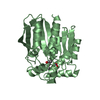
| ||||||||
| Unit cell |
| ||||||||
| Components on special symmetry positions |
|
-Components
| #1: Protein | Mass: 30298.533 Da / Num. of mol.: 3 Source method: isolated from a genetically manipulated source Source: (gene. exp.) Mycobacterium tuberculosis H37Rv (bacteria)Gene: Rv0183 / Production host: #2: Chemical | ChemComp-SO4 / |   Mass: 96.063 Da / Num. of mol.: 1 / Source method: obtained synthetically / Formula: SO4 Mass: 96.063 Da / Num. of mol.: 1 / Source method: obtained synthetically / Formula: SO4#3: Chemical |   Mass: 118.174 Da / Num. of mol.: 3 / Source method: obtained synthetically / Formula: C6H14O2 / Comment: precipitant*YM Mass: 118.174 Da / Num. of mol.: 3 / Source method: obtained synthetically / Formula: C6H14O2 / Comment: precipitant*YM#4: Chemical | ChemComp-NO3 / |   Mass: 62.005 Da / Num. of mol.: 1 / Source method: obtained synthetically / Formula: NO3 Mass: 62.005 Da / Num. of mol.: 1 / Source method: obtained synthetically / Formula: NO3#5: Water | ChemComp-HOH / |  Mass: 18.015 Da / Num. of mol.: 459 / Source method: isolated from a natural source / Formula: H2O Mass: 18.015 Da / Num. of mol.: 459 / Source method: isolated from a natural source / Formula: H2O |
|---|
-Experimental details
-Experiment
| Experiment | Method: X-RAY DIFFRACTION / Number of used crystals: 1 |
|---|
- Sample preparation
| Crystal | Density Matthews: 2.33 Å3/Da / Density % sol: 47.32 % |
|---|---|
| Crystal grow | Temperature: 293.15 K / Method: vapor diffusion, sitting drop / pH: 7.8 Details: 0.03M sodium nitrate, 0.03M di-sodium hydrogenphosphate, 0.03M ammonium sulfate, 0.1M MOPS/HEPES, 12% 2-Methyl 2,4 pentandiol, 12% PEG1000, 12% PEG3350. Microseeding |
-Data collection
| Diffraction | Mean temperature: 100 K |
|---|---|
| Diffraction source | Source: SYNCHROTRON / Site: ESRF  / Beamline: ID23-1 / Wavelength: 0.97895 Å / Beamline: ID23-1 / Wavelength: 0.97895 Å |
| Detector | Type: DECTRIS PILATUS 6M / Detector: PIXEL / Date: Mar 1, 2015 |
| Radiation | Protocol: SINGLE WAVELENGTH / Monochromatic (M) / Laue (L): M / Scattering type: x-ray |
| Radiation wavelength | Wavelength: 0.97895 Å / Relative weight: 1 |
| Reflection | Resolution: 1.8→49.209 Å / Num. obs: 77304 / % possible obs: 100 % / Redundancy: 9.6 % / CC1/2: 0.998 / Rmerge(I) obs: 0.105 / Rpim(I) all: 0.035 / Net I/σ(I): 16.4 |
| Reflection shell | Resolution: 1.8→1.84 Å / Redundancy: 8.7 % / Rmerge(I) obs: 0.853 / Mean I/σ(I) obs: 2.6 / Num. unique obs: 4549 / CC1/2: 0.734 / Rpim(I) all: 0.307 / % possible all: 100 |
- Processing
| Software |
| |||||||||||||||||||||||||||||||||||||||||||||||||||||||||||||||||||||||||||||||||||||||||||||||||||||||||||||||||||||||||||||||||||||||||||||||||||
|---|---|---|---|---|---|---|---|---|---|---|---|---|---|---|---|---|---|---|---|---|---|---|---|---|---|---|---|---|---|---|---|---|---|---|---|---|---|---|---|---|---|---|---|---|---|---|---|---|---|---|---|---|---|---|---|---|---|---|---|---|---|---|---|---|---|---|---|---|---|---|---|---|---|---|---|---|---|---|---|---|---|---|---|---|---|---|---|---|---|---|---|---|---|---|---|---|---|---|---|---|---|---|---|---|---|---|---|---|---|---|---|---|---|---|---|---|---|---|---|---|---|---|---|---|---|---|---|---|---|---|---|---|---|---|---|---|---|---|---|---|---|---|---|---|---|---|---|---|
| Refinement | Method to determine structure: MOLECULAR REPLACEMENT / Resolution: 1.8→49.209 Å / Cross valid method: FREE R-VALUE / σ(F): 1.35 / Phase error: 28.91
| |||||||||||||||||||||||||||||||||||||||||||||||||||||||||||||||||||||||||||||||||||||||||||||||||||||||||||||||||||||||||||||||||||||||||||||||||||
| Solvent computation | Shrinkage radii: 0.9 Å / VDW probe radii: 1.11 Å | |||||||||||||||||||||||||||||||||||||||||||||||||||||||||||||||||||||||||||||||||||||||||||||||||||||||||||||||||||||||||||||||||||||||||||||||||||
| Refinement step | Cycle: LAST / Resolution: 1.8→49.209 Å
| |||||||||||||||||||||||||||||||||||||||||||||||||||||||||||||||||||||||||||||||||||||||||||||||||||||||||||||||||||||||||||||||||||||||||||||||||||
| Refine LS restraints |
| |||||||||||||||||||||||||||||||||||||||||||||||||||||||||||||||||||||||||||||||||||||||||||||||||||||||||||||||||||||||||||||||||||||||||||||||||||
| LS refinement shell |
|
