 Movie
Movie Controller


+ Open data

- Basic information
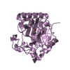
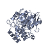
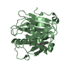
| Entry | Database: PDB / ID: 1va4 | ||||||
|---|---|---|---|---|---|---|---|
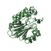
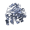
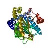
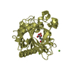
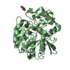
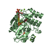
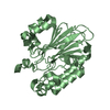
| Title | Pseudomonas fluorescens aryl esterase | ||||||
 Components Components | Arylesterase | ||||||
 Keywords Keywords | HYDROLASE / alpha/beta hydrolase / esterase / non-cofactor dependent haloperoxidase | ||||||
| Function / homology |  Function and homology information Function and homology informationarylesterase / Oxidoreductases / arylesterase activity / peroxidase activity Similarity search - Function | ||||||
| Biological species |  Pseudomonas fluorescens (bacteria) Pseudomonas fluorescens (bacteria) | ||||||
| Method |  X-RAY DIFFRACTION / MOLECULAR REPLACEMENT / Resolution: 1.804 Å X-RAY DIFFRACTION / MOLECULAR REPLACEMENT / Resolution: 1.804 Å | ||||||
 Authors Authors | Cheeseman, J.D. / Tocilj, A. / Park, S. / Schrag, J.D. / Kazlauskas, R.J. | ||||||
 Citation Citation | Journal: Acta Crystallogr.,Sect.D / Year: 2004 Title: Structure of an aryl esterase from Pseudomonas fluorescens. Authors: Cheeseman, J.D. / Tocilj, A. / Park, S. / Schrag, J.D. / Kazlauskas, R.J. | ||||||
| History |
|
- Structure visualization
| Structure viewer | Molecule: MolmilJmol/JSmol |
|---|
- Downloads & links
-Download
| PDBx/mmCIF format | 1va4.cif.gz | 349.5 KB | Display | PDBx/mmCIF format |
|---|---|---|---|---|
| PDB format | pdb1va4.ent.gz | 284.4 KB | Display | PDB format |
| PDBx/mmJSON format | 1va4.json.gz | Tree view | PDBx/mmJSON format | |
| Others |  Other downloads Other downloads |
-Validation report
| Arichive directory | https://data.pdbj.org/pub/pdb/validation_reports/va/1va4ftp://data.pdbj.org/pub/pdb/validation_reports/va/1va4 | HTTPS FTP |
|---|
-Related structure data
| Related structure data | 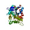 1a8sS S: Starting model for refinement |
|---|---|
| Similar structure data |
-Links
 PDBj
PDBj


- Assembly
| Deposited unit | 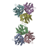
| ||||||||
|---|---|---|---|---|---|---|---|---|---|
| 1 | 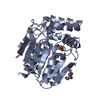
| ||||||||
| 2 | 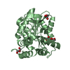
| ||||||||
| 3 | 
| ||||||||
| 4 | 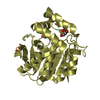
| ||||||||
| 5 | 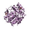
| ||||||||
| 6 | 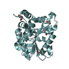
| ||||||||
| Unit cell |
|
-Components
| #1: Protein | Mass: 30966.955 Da / Num. of mol.: 6 Source method: isolated from a genetically manipulated source Source: (gene. exp.) Pseudomonas fluorescens (bacteria) / Strain: SIK WI / Plasmid: pUE1251 / Production host: #2: Chemical | ChemComp-GOL /   Mass: 92.094 Da / Num. of mol.: 20 / Source method: obtained synthetically / Formula: C3H8O3 Mass: 92.094 Da / Num. of mol.: 20 / Source method: obtained synthetically / Formula: C3H8O3#3: Water | ChemComp-HOH / |  Mass: 18.015 Da / Num. of mol.: 1362 / Source method: isolated from a natural source / Formula: H2O Mass: 18.015 Da / Num. of mol.: 1362 / Source method: isolated from a natural source / Formula: H2O |
|---|
-Experimental details
-Experiment
| Experiment | Method: X-RAY DIFFRACTION / Number of used crystals: 1 |
|---|
- Sample preparation
| Crystal | Density Matthews: 4.3 Å3/Da / Density % sol: 71.41 % |
|---|---|
| Crystal grow | Temperature: 293 K / Method: vapor diffusion, hanging drop / pH: 7.5 Details: 1% PEG 400, 1.65M (NH4)2SO4, 0.1M HEPES , pH 7.5, VAPOR DIFFUSION, HANGING DROP, temperature 293K |
-Data collection
| Diffraction | Mean temperature: 93 K |
|---|---|
| Diffraction source | Source: ROTATING ANODE / Type: RIGAKU MICROMAX-007 / Wavelength: 1.5418 Å |
| Detector | Type: RIGAKU RAXIS HTC / Detector: IMAGE PLATE / Date: Apr 15, 2003 / Details: Osmic mirrors |
| Radiation | Protocol: SINGLE WAVELENGTH / Monochromatic (M) / Laue (L): M / Scattering type: x-ray |
| Radiation wavelength | Wavelength: 1.5418 Å / Relative weight: 1 |
| Reflection | Resolution: 1.8→48.8 Å / Num. all: 285139 / Num. obs: 271452 / % possible obs: 95.2 % / Observed criterion σ(F): 1 / Observed criterion σ(I): 1 / Redundancy: 5.04 % / Rsym value: 0.066 / Net I/σ(I): 14 |
| Reflection shell | Resolution: 1.804→1.851 Å / Redundancy: 2.43 % / Mean I/σ(I) obs: 3.7 / Num. unique all: 1314 / Rsym value: 0.247 / % possible all: 65.8 |
- Processing
| Software |
| ||||||||||||||||||||||||||||||||||||||||||||||||||||||||||||||||||||||
|---|---|---|---|---|---|---|---|---|---|---|---|---|---|---|---|---|---|---|---|---|---|---|---|---|---|---|---|---|---|---|---|---|---|---|---|---|---|---|---|---|---|---|---|---|---|---|---|---|---|---|---|---|---|---|---|---|---|---|---|---|---|---|---|---|---|---|---|---|---|---|---|
| Refinement | Method to determine structure: MOLECULAR REPLACEMENT Starting model: 1A8S Resolution: 1.804→48.53 Å / Cor.coef. Fo:Fc: 0.959 / Cor.coef. Fo:Fc free: 0.948 / SU B: 1.77 / SU ML: 0.052 / Isotropic thermal model: Isotropic / Cross valid method: THROUGHOUT / σ(F): 0 / ESU R: 0.089 / ESU R Free: 0.09 / Stereochemistry target values: MAXIMUM LIKELIHOOD
| ||||||||||||||||||||||||||||||||||||||||||||||||||||||||||||||||||||||
| Solvent computation | Ion probe radii: 0.8 Å / Shrinkage radii: 0.8 Å / VDW probe radii: 1.4 Å / Solvent model: BABINET MODEL WITH MASK | ||||||||||||||||||||||||||||||||||||||||||||||||||||||||||||||||||||||
| Displacement parameters | Biso mean: 18.672 Å2
| ||||||||||||||||||||||||||||||||||||||||||||||||||||||||||||||||||||||
| Refinement step | Cycle: LAST / Resolution: 1.804→48.53 Å
| ||||||||||||||||||||||||||||||||||||||||||||||||||||||||||||||||||||||
| Refine LS restraints |
| ||||||||||||||||||||||||||||||||||||||||||||||||||||||||||||||||||||||
| LS refinement shell | Resolution: 1.804→1.851 Å / Total num. of bins used: 20 /
|
