 Movie
Movie Controller

[English] 日本語
 Yorodumi
- PDB-5dm4: Crystal structure of the plantazolicin methyltransferase BpumL in... -
+ Open data

- Basic information
| Entry | Database: PDB / ID: 5dm4 | ||||||
|---|---|---|---|---|---|---|---|
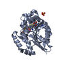
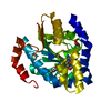
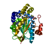
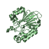
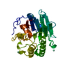
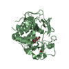
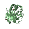
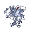
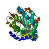
| Title | Crystal structure of the plantazolicin methyltransferase BpumL in complex with pentazolic desmethylPZN analog and SAH | ||||||
 Components Components | Methyltransferase domain family | ||||||
 Keywords Keywords | transferase/transferase inhibitor / N-methyltransferase / plantazolicin / pentazolic desmethylPZN analog / transferase-transferase inhibitor complex | ||||||
| Function / homology | Vaccinia Virus protein VP39 / Rossmann fold / 3-Layer(aba) Sandwich / Alpha Beta / Chem-5DA / S-ADENOSYL-L-HOMOCYSTEINE / :  Function and homology information Function and homology information | ||||||
| Biological species |  | ||||||
| Method |  X-RAY DIFFRACTION / SYNCHROTRON / Resolution: 1.75 Å X-RAY DIFFRACTION / SYNCHROTRON / Resolution: 1.75 Å | ||||||
 Authors Authors | Hao, Y. / Nair, S.K. | ||||||
 Citation Citation | Journal: Acs Chem.Biol. / Year: 2015 Title: Insights into methyltransferase specificity and bioactivity of derivatives of the antibiotic plantazolicin. Authors: Hao, Y. / Blair, P.M. / Sharma, A. / Mitchell, D.A. / Nair, S.K. | ||||||
| History |
|
- Structure visualization
| Structure viewer | Molecule: MolmilJmol/JSmol |
|---|
- Downloads & links
-Download
| PDBx/mmCIF format | 5dm4.cif.gz | 72.8 KB | Display | PDBx/mmCIF format |
|---|---|---|---|---|
| PDB format | pdb5dm4.ent.gz | 52 KB | Display | PDB format |
| PDBx/mmJSON format | 5dm4.json.gz | Tree view | PDBx/mmJSON format | |
| Others |  Other downloads Other downloads |
-Validation report
| Arichive directory | https://data.pdbj.org/pub/pdb/validation_reports/dm/5dm4ftp://data.pdbj.org/pub/pdb/validation_reports/dm/5dm4 | HTTPS FTP |
|---|
-Related structure data


-Links
 PDBj
PDBj- Assembly
| Deposited unit | 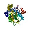
| ||||||||
|---|---|---|---|---|---|---|---|---|---|
| 1 |
| ||||||||
| Unit cell |
|
-Components
| #1: Protein | Mass: 30385.580 Da / Num. of mol.: 1 Source method: isolated from a genetically manipulated source Source: (gene. exp.) |
|---|---|
| #2: Chemical | ChemComp-SAH /   Type: L-peptide linking / Mass: 384.411 Da / Num. of mol.: 1 Type: L-peptide linking / Mass: 384.411 Da / Num. of mol.: 1Source method: isolated from a genetically manipulated source Formula: C14H20N6O5S |
| #3: Chemical | ChemComp-GOL /   Mass: 92.094 Da / Num. of mol.: 1 Mass: 92.094 Da / Num. of mol.: 1Source method: isolated from a genetically manipulated source Formula: C3H8O3 |
| #4: Chemical | ChemComp-5DA /   Mass: 553.660 Da / Num. of mol.: 1 Mass: 553.660 Da / Num. of mol.: 1Source method: isolated from a genetically manipulated source Formula: C24H27N9O3S2 |
| #5: Water | ChemComp-HOH /  Mass: 18.015 Da / Num. of mol.: 186 / Source method: isolated from a natural source / Formula: H2O Mass: 18.015 Da / Num. of mol.: 186 / Source method: isolated from a natural source / Formula: H2O |
-Experimental details
-Experiment
| Experiment | Method: X-RAY DIFFRACTION |
|---|
- Sample preparation
| Crystal | Density Matthews: 1.96 Å3/Da / Density % sol: 37.19 % |
|---|---|
| Crystal grow | Temperature: 282 K / Method: vapor diffusion, hanging drop Details: 100mM Bis-tris pH 5.5, 200mM Li2SO4, 22-26% PEG3350 |
-Data collection
| Diffraction | Mean temperature: 80 K |
|---|---|
| Diffraction source | Source: SYNCHROTRON / Site: APS  / Beamline: 21-ID-G / Wavelength: 0.987 Å / Beamline: 21-ID-G / Wavelength: 0.987 Å |
| Detector | Type: RAYONIX MX-300 / Detector: CCD / Date: Jul 28, 2013 |
| Radiation | Protocol: SINGLE WAVELENGTH / Monochromatic (M) / Laue (L): M / Scattering type: x-ray |
| Radiation wavelength | Wavelength: 0.987 Å / Relative weight: 1 |
| Reflection | Resolution: 1.75→25 Å / Num. obs: 25053 / % possible obs: 99.96 % / Redundancy: 8 % / Net I/σ(I): 10.7 |
- Processing
| Software |
| ||||||||||||||||||||||||||||||||||||||||||||||||||||||||||||||||||||||||||||||||||||||||||||||||||||||||||||||||||||||||||||||||||||||||||||||||||||||||||||||||||||||||||||||||||||||
|---|---|---|---|---|---|---|---|---|---|---|---|---|---|---|---|---|---|---|---|---|---|---|---|---|---|---|---|---|---|---|---|---|---|---|---|---|---|---|---|---|---|---|---|---|---|---|---|---|---|---|---|---|---|---|---|---|---|---|---|---|---|---|---|---|---|---|---|---|---|---|---|---|---|---|---|---|---|---|---|---|---|---|---|---|---|---|---|---|---|---|---|---|---|---|---|---|---|---|---|---|---|---|---|---|---|---|---|---|---|---|---|---|---|---|---|---|---|---|---|---|---|---|---|---|---|---|---|---|---|---|---|---|---|---|---|---|---|---|---|---|---|---|---|---|---|---|---|---|---|---|---|---|---|---|---|---|---|---|---|---|---|---|---|---|---|---|---|---|---|---|---|---|---|---|---|---|---|---|---|---|---|---|---|
| Refinement | Resolution: 1.75→25 Å / Cor.coef. Fo:Fc: 0.95 / Cor.coef. Fo:Fc free: 0.925 / SU B: 2.633 / SU ML: 0.087 / Cross valid method: THROUGHOUT / ESU R: 0.144 / ESU R Free: 0.136 / Stereochemistry target values: MAXIMUM LIKELIHOOD / Details: HYDROGENS HAVE BEEN USED IF PRESENT IN THE INPUT
| ||||||||||||||||||||||||||||||||||||||||||||||||||||||||||||||||||||||||||||||||||||||||||||||||||||||||||||||||||||||||||||||||||||||||||||||||||||||||||||||||||||||||||||||||||||||
| Solvent computation | Ion probe radii: 0.8 Å / Shrinkage radii: 0.8 Å / VDW probe radii: 1.2 Å / Solvent model: BABINET MODEL WITH MASK | ||||||||||||||||||||||||||||||||||||||||||||||||||||||||||||||||||||||||||||||||||||||||||||||||||||||||||||||||||||||||||||||||||||||||||||||||||||||||||||||||||||||||||||||||||||||
| Displacement parameters | Biso mean: 20.6 Å2
| ||||||||||||||||||||||||||||||||||||||||||||||||||||||||||||||||||||||||||||||||||||||||||||||||||||||||||||||||||||||||||||||||||||||||||||||||||||||||||||||||||||||||||||||||||||||
| Refinement step | Cycle: 1 / Resolution: 1.75→25 Å
| ||||||||||||||||||||||||||||||||||||||||||||||||||||||||||||||||||||||||||||||||||||||||||||||||||||||||||||||||||||||||||||||||||||||||||||||||||||||||||||||||||||||||||||||||||||||
| Refine LS restraints |
|