Monochromator: double crystal / Protocol: SINGLE WAVELENGTH / Monochromatic (M) / Laue (L): M / Scattering type: x-ray
Radiation wavelength
Wavelength: 0.9195 Å / Relative weight: 1
Reflection
Resolution: 1.8→42.18 Å / Num. all: 57215 / Num. obs: 57215 / % possible obs: 95.3 % / Redundancy: 7.6 % / Biso Wilson estimate: 17.2 Å2 / Rsym value: 0.072 / Net I/σ(I): 18.2
Reflection shell
Resolution: 1.8→1.9 Å / Redundancy: 5.9 % / Mean I/σ(I) obs: 2.4 / Num. unique all: 7474 / Rsym value: 0.337 / % possible all: 86.4
-
Processing
Software
Name
Version
Classification
NB
SCALA
datascaling
REFMAC
5.2.0005
refinement
PDB_EXTRACT
1.701
dataextraction
MOSFLM
datareduction
CCP4
(SCALA)
datascaling
CNS
phasing
Refinement
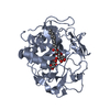
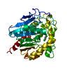
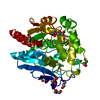
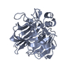
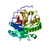
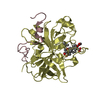
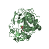
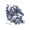
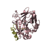
Method to determine structure: RIGID BODY Starting model: SyrB2 with Fe(II), chloride, and alpha-ketoglutarate Resolution: 1.8→42.18 Å / Cor.coef. Fo:Fc: 0.955 / Cor.coef. Fo:Fc free: 0.938 / SU B: 2.307 / SU ML: 0.074 / Cross valid method: THROUGHOUT / ESU R: 0.131 / ESU R Free: 0.125 / Stereochemistry target values: MAXIMUM LIKELIHOOD / Details: HYDROGENS HAVE BEEN ADDED IN THE RIDING POSITIONS
Rfactor
Num. reflection
% reflection
Selection details
Rfree
0.21
3341
5.9 %
Same as starting model
Rwork
0.171
-
-
-
all
0.173
56498
-
-
obs
0.173
56498
93.98 %
-
Solvent computation
Ion probe radii: 0.8 Å / Shrinkage radii: 0.8 Å / VDW probe radii: 1.2 Å / Solvent model: MASK
Displacement parameters
Biso mean: 17.096 Å2
Baniso -1
Baniso -2
Baniso -3
1-
-0.23 Å2
0 Å2
0 Å2
2-
-
-0.11 Å2
0 Å2
3-
-
-
0.35 Å2
Refine analyze
Luzzati coordinate error obs: 0.183 Å
Refinement step
Cycle: LAST / Resolution: 1.8→42.18 Å
Protein
Nucleic acid
Ligand
Solvent
Total
Num. atoms
4814
0
60
465
5339
Refine LS restraints
Refine-ID
Type
Dev ideal
Dev ideal target
Number
X-RAY DIFFRACTION
r_bond_refined_d
0.015
0.022
5020
X-RAY DIFFRACTION
r_bond_other_d
0.002
0.02
4267
X-RAY DIFFRACTION
r_angle_refined_deg
1.532
1.934
6811
X-RAY DIFFRACTION
r_angle_other_deg
1.345
3
9943
X-RAY DIFFRACTION
r_dihedral_angle_1_deg
6.836
5
599
X-RAY DIFFRACTION
r_dihedral_angle_2_deg
32.644
23.91
266
X-RAY DIFFRACTION
r_dihedral_angle_3_deg
14.486
15
768
X-RAY DIFFRACTION
r_dihedral_angle_4_deg
13.923
15
34
X-RAY DIFFRACTION
r_chiral_restr
0.097
0.2
694
X-RAY DIFFRACTION
r_gen_planes_refined
0.007
0.02
5687
X-RAY DIFFRACTION
r_gen_planes_other
0.001
0.02
1097
X-RAY DIFFRACTION
r_nbd_refined
0.208
0.2
926
X-RAY DIFFRACTION
r_nbd_other
0.203
0.2
4267
X-RAY DIFFRACTION
r_nbtor_refined
0.187
0.2
2384
X-RAY DIFFRACTION
r_nbtor_other
0.089
0.2
2572
X-RAY DIFFRACTION
r_xyhbond_nbd_refined
0.152
0.2
361
X-RAY DIFFRACTION
r_symmetry_vdw_refined
0.17
0.2
10
X-RAY DIFFRACTION
r_symmetry_vdw_other
0.282
0.2
39
X-RAY DIFFRACTION
r_symmetry_hbond_refined
0.16
0.2
12
X-RAY DIFFRACTION
r_mcbond_it
1.387
1.5
3819
X-RAY DIFFRACTION
r_mcbond_other
0.252
1.5
1220
X-RAY DIFFRACTION
r_mcangle_it
1.535
2
4823
X-RAY DIFFRACTION
r_scbond_it
2.474
3
2422
X-RAY DIFFRACTION
r_scangle_it
3.437
4.5
1988
LS refinement shell
Resolution: 1.8→1.847 Å / Total num. of bins used: 20
Rfactor
Num. reflection
% reflection
Rfree
0.249
296
-
Rwork
0.201
3309
-
obs
-
3605
82.01 %
+
About Yorodumi
-
News
-
Feb 9, 2022. New format data for meta-information of EMDB entries
New format data for meta-information of EMDB entries
Version 3 of the EMDB header file is now the official format.
The previous official version 1.9 will be removed from the archive.
In the structure databanks used in Yorodumi, some data are registered as the other names, "COVID-19 virus" and "2019-nCoV". Here are the details of the virus and the list of structure data.
Jan 31, 2019. EMDB accession codes are about to change! (news from PDBe EMDB page)
EMDB accession codes are about to change! (news from PDBe EMDB page)
The allocation of 4 digits for EMDB accession codes will soon come to an end. Whilst these codes will remain in use, new EMDB accession codes will include an additional digit and will expand incrementally as the available range of codes is exhausted. The current 4-digit format prefixed with “EMD-” (i.e. EMD-XXXX) will advance to a 5-digit format (i.e. EMD-XXXXX), and so on. It is currently estimated that the 4-digit codes will be depleted around Spring 2019, at which point the 5-digit format will come into force.
The EM Navigator/Yorodumi systems omit the EMD- prefix.
Related info.:Q: What is EMD? / ID/Accession-code notation in Yorodumi/EM Navigator
Yorodumi is a browser for structure data from EMDB, PDB, SASBDB, etc.
This page is also the successor to EM Navigator detail page, and also detail information page/front-end page for Omokage search.
The word "yorodu" (or yorozu) is an old Japanese word meaning "ten thousand". "mi" (miru) is to see.
Related info.:EMDB / PDB / SASBDB / Comparison of 3 databanks / Yorodumi Search / Aug 31, 2016. New EM Navigator & Yorodumi / Yorodumi Papers / Jmol/JSmol / Function and homology information / Changes in new EM Navigator and Yorodumi
 Movie
Movie Controller


 Open data
 Basic information
 Components
 Keywords
 Function and homology information
 Pseudomonas syringae (bacteria)
 X-RAY DIFFRACTION /
  Authors
 Citation
 Structure visualization
 Downloads & links
 Other downloads
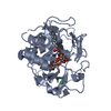


 PDBj
PDBj


 Assembly
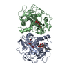

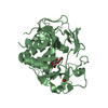


 Mass: 55.845 Da / Num. of mol.: 2 / Source method: obtained synthetically / Formula: Fe
 Mass: 79.904 Da / Num. of mol.: 4 / Source method: obtained synthetically / Formula: Br
 Mass: 496.546 Da / Num. of mol.: 1 / Source method: obtained synthetically / Formula: C22H40O12
 Mass: 146.098 Da / Num. of mol.: 2 / Source method: obtained synthetically / Formula: C5H6O5
 Sample preparation
 / Beamline: BL9-2 / Wavelength: 0.9195 Å
 Processing