- PDB-6mk3: Crystallographic solvent mapping analysis of DMSO bound to APE1 -
+
Open data
ID or keywords:
Loading...
-
Basic information
Entry
Database: PDB / ID: 6mk3
Title
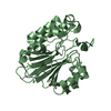
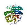
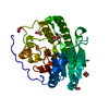
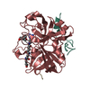
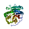
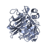
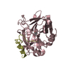
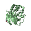
Crystallographic solvent mapping analysis of DMSO bound to APE1
Components
DNA-(apurinic or apyrimidinic site) lyase
Keywords
LYASE / Apurinic/apyrimidinic endonuclease / DNA repair / abasic site / solvent mapping
Function / homology
Function and homology information
Resolution of Abasic Sites (AP sites) / phosphodiesterase activity, acting on 3'-phosphoglycolate-terminated DNA strands / telomere maintenance via base-excision repair / class II DNA-(apurinic or apyrimidinic site) endonuclease activity / deoxyribonuclease (pyrimidine dimer) activity / DNA-(abasic site) binding / double-stranded DNA exodeoxyribonuclease activity / exodeoxyribonuclease III / double-stranded DNA 3'-5' DNA exonuclease activity / Displacement of DNA glycosylase by APEX1 ...Resolution of Abasic Sites (AP sites) / phosphodiesterase activity, acting on 3'-phosphoglycolate-terminated DNA strands / telomere maintenance via base-excision repair / class II DNA-(apurinic or apyrimidinic site) endonuclease activity / deoxyribonuclease (pyrimidine dimer) activity / DNA-(abasic site) binding / double-stranded DNA exodeoxyribonuclease activity / exodeoxyribonuclease III / double-stranded DNA 3'-5' DNA exonuclease activity / Displacement of DNA glycosylase by APEX1 / double-stranded telomeric DNA binding / positive regulation of gene expression via chromosomal CpG island demethylation / uracil DNA N-glycosylase activity / phosphoric diester hydrolase activity / 3'-5'-DNA exonuclease activity / Hydrolases; Acting on ester bonds; Endodeoxyribonucleases producing 5'-phosphomonoesters / phosphodiesterase I activity / DNA catabolic process / Resolution of AP sites via the multiple-nucleotide patch replacement pathway / Abasic sugar-phosphate removal via the single-nucleotide replacement pathway / POLB-Dependent Long Patch Base Excision Repair / PCNA-Dependent Long Patch Base Excision Repair / base-excision repair, gap-filling / 3'-5' exonuclease activity / regulation of mRNA stability / DNA-(apurinic or apyrimidinic site) endonuclease activity / cell redox homeostasis / telomere maintenance / DNA endonuclease activity / chromatin DNA binding / base-excision repair / RNA-DNA hybrid ribonuclease activity / transcription corepressor activity / endonuclease activity / DNA recombination / regulation of apoptotic process / damaged DNA binding / transcription coactivator activity / chromosome, telomeric region / oxidoreductase activity / nuclear speck / ribosome / DNA repair / centrosome / nucleolus / perinuclear region of cytoplasm / endoplasmic reticulum / positive regulation of transcription by RNA polymerase II / mitochondrion / DNA binding / RNA binding / nucleoplasm / metal ion binding / nucleus / cytoplasm Similarity search - Function
AP endonucleases family 1 signature 2. / AP endonuclease 1, conserved site / AP endonucleases family 1 signature 3. / AP endonuclease 1, binding site / AP endonucleases family 1 signature 1. / AP endonuclease 1 / AP endonucleases family 1 profile. / Deoxyribonuclease I; Chain A / Endonuclease/exonuclease/phosphatase / Endonuclease/exonuclease/phosphatase ...AP endonucleases family 1 signature 2. / AP endonuclease 1, conserved site / AP endonucleases family 1 signature 3. / AP endonuclease 1, binding site / AP endonucleases family 1 signature 1. / AP endonuclease 1 / AP endonucleases family 1 profile. / Deoxyribonuclease I; Chain A / Endonuclease/exonuclease/phosphatase / Endonuclease/exonuclease/phosphatase / Endonuclease/Exonuclease/phosphatase family / Endonuclease/exonuclease/phosphatase superfamily / 4-Layer Sandwich / Alpha Beta Similarity search - Domain/homology
In the structure databanks used in Yorodumi, some data are registered as the other names, "COVID-19 virus" and "2019-nCoV". Here are the details of the virus and the list of structure data.
Jan 31, 2019. EMDB accession codes are about to change! (news from PDBe EMDB page)
EMDB accession codes are about to change! (news from PDBe EMDB page)
The allocation of 4 digits for EMDB accession codes will soon come to an end. Whilst these codes will remain in use, new EMDB accession codes will include an additional digit and will expand incrementally as the available range of codes is exhausted. The current 4-digit format prefixed with “EMD-” (i.e. EMD-XXXX) will advance to a 5-digit format (i.e. EMD-XXXXX), and so on. It is currently estimated that the 4-digit codes will be depleted around Spring 2019, at which point the 5-digit format will come into force.
The EM Navigator/Yorodumi systems omit the EMD- prefix.
Related info.:Q: What is EMD? / ID/Accession-code notation in Yorodumi/EM Navigator
Yorodumi is a browser for structure data from EMDB, PDB, SASBDB, etc.
This page is also the successor to EM Navigator detail page, and also detail information page/front-end page for Omokage search.
The word "yorodu" (or yorozu) is an old Japanese word meaning "ten thousand". "mi" (miru) is to see.
Related info.:EMDB / PDB / SASBDB / Comparison of 3 databanks / Yorodumi Search / Aug 31, 2016. New EM Navigator & Yorodumi / Yorodumi Papers / Jmol/JSmol / Function and homology information / Changes in new EM Navigator and Yorodumi
 Movie
Movie Controller


 Open data
 Basic information
 Components
 Keywords
 Function and homology information
 Homo sapiens (human)
 X-RAY DIFFRACTION /
  Authors
 United States, 1items
  Citation
 Structure visualization
 Downloads & links
 Other downloads


 PDBj
PDBj


 Assembly


 Mass: 78.133 Da / Num. of mol.: 2 / Source method: obtained synthetically / Formula: C2H6OS / Comment: DMSO, precipitant*YM
 Mass: 62.068 Da / Num. of mol.: 3 / Source method: obtained synthetically / Formula: C2H6O2
 Mass: 18.015 Da / Num. of mol.: 252 / Source method: isolated from a natural source / Formula: H2O
 Sample preparation
 Processing