 Movie
Movie Controller


+ Open data

- Basic information
| Entry | Database: PDB / ID: 5wn3 | ||||||
|---|---|---|---|---|---|---|---|
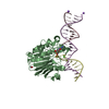
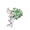
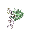
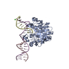
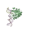
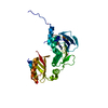
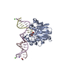
| Title | APE1 F266A exonuclease substrate complex with a C/T mismatch | ||||||
 Components Components |
| ||||||
 Keywords Keywords | HYDROLASE / LYASE/DNA / LYASE-DNA complex | ||||||
| Function / homology |  Function and homology information Function and homology informationResolution of Abasic Sites (AP sites) / phosphodiesterase activity, acting on 3'-phosphoglycolate-terminated DNA strands / telomere maintenance via base-excision repair / class II DNA-(apurinic or apyrimidinic site) endonuclease activity / deoxyribonuclease (pyrimidine dimer) activity / DNA-(abasic site) binding / double-stranded DNA exodeoxyribonuclease activity / exodeoxyribonuclease III / double-stranded DNA 3'-5' DNA exonuclease activity / Displacement of DNA glycosylase by APEX1 ...Resolution of Abasic Sites (AP sites) / phosphodiesterase activity, acting on 3'-phosphoglycolate-terminated DNA strands / telomere maintenance via base-excision repair / class II DNA-(apurinic or apyrimidinic site) endonuclease activity / deoxyribonuclease (pyrimidine dimer) activity / DNA-(abasic site) binding / double-stranded DNA exodeoxyribonuclease activity / exodeoxyribonuclease III / double-stranded DNA 3'-5' DNA exonuclease activity / Displacement of DNA glycosylase by APEX1 / double-stranded telomeric DNA binding / positive regulation of gene expression via chromosomal CpG island demethylation / uracil DNA N-glycosylase activity / phosphoric diester hydrolase activity / 3'-5'-DNA exonuclease activity / Hydrolases; Acting on ester bonds; Endodeoxyribonucleases producing 5'-phosphomonoesters / phosphodiesterase I activity / DNA catabolic process / Resolution of AP sites via the multiple-nucleotide patch replacement pathway / Abasic sugar-phosphate removal via the single-nucleotide replacement pathway / POLB-Dependent Long Patch Base Excision Repair / PCNA-Dependent Long Patch Base Excision Repair / base-excision repair, gap-filling / 3'-5' exonuclease activity / regulation of mRNA stability / DNA-(apurinic or apyrimidinic site) endonuclease activity / cell redox homeostasis / telomere maintenance / DNA endonuclease activity / chromatin DNA binding / base-excision repair / RNA-DNA hybrid ribonuclease activity / transcription corepressor activity / endonuclease activity / DNA recombination / regulation of apoptotic process / damaged DNA binding / transcription coactivator activity / chromosome, telomeric region / oxidoreductase activity / nuclear speck / ribosome / DNA repair / centrosome / nucleolus / perinuclear region of cytoplasm / endoplasmic reticulum / positive regulation of transcription by RNA polymerase II / mitochondrion / DNA binding / RNA binding / nucleoplasm / metal ion binding / nucleus / cytoplasm Similarity search - Function | ||||||
| Biological species |  Homo sapiens (human) Homo sapiens (human)synthetic construct (others) | ||||||
| Method |  X-RAY DIFFRACTION / MOLECULAR REPLACEMENT / Resolution: 2 Å X-RAY DIFFRACTION / MOLECULAR REPLACEMENT / Resolution: 2 Å | ||||||
 Authors Authors | Freudenthal, B.D. / Whitaker, A.M. | ||||||
| Funding support |  United States, 1items United States, 1items
| ||||||
 Citation Citation | Journal: Nat Commun / Year: 2018 Title: Molecular snapshots of APE1 proofreading mismatches and removing DNA damage. Authors: Whitaker, A.M. / Flynn, T.S. / Freudenthal, B.D. | ||||||
| History |
|
- Structure visualization
| Structure viewer | Molecule: MolmilJmol/JSmol |
|---|
- Downloads & links
-Download
| PDBx/mmCIF format | 5wn3.cif.gz | 156.3 KB | Display | PDBx/mmCIF format |
|---|---|---|---|---|
| PDB format | pdb5wn3.ent.gz | 118.1 KB | Display | PDB format |
| PDBx/mmJSON format | 5wn3.json.gz | Tree view | PDBx/mmJSON format | |
| Others |  Other downloads Other downloads |
-Validation report
| Arichive directory | https://data.pdbj.org/pub/pdb/validation_reports/wn/5wn3ftp://data.pdbj.org/pub/pdb/validation_reports/wn/5wn3 | HTTPS FTP |
|---|
-Related structure data
| Related structure data |  5wn0C  5wn1C  5wn2C  5wn4C  5wn5C 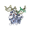 5dffS S: Starting model for refinement C: citing same article ( |
|---|---|
| Similar structure data |


-Links
 PDBj
PDBj


- Assembly
| Deposited unit | 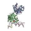
| ||||||||
|---|---|---|---|---|---|---|---|---|---|
| 1 | 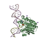
| ||||||||
| 2 | 
| ||||||||
| Unit cell |
|
-Components
-DNA chain , 3 types, 3 molecules CDE
| #1: DNA chain | Mass: 3334.186 Da / Num. of mol.: 1 / Source method: obtained synthetically / Source: (synth.) synthetic construct (others) |
|---|---|
| #2: DNA chain | Mass: 3077.070 Da / Num. of mol.: 1 / Source method: obtained synthetically / Source: (synth.) synthetic construct (others) |
| #3: DNA chain | Mass: 6424.148 Da / Num. of mol.: 1 / Source method: obtained synthetically / Source: (synth.) synthetic construct (others) |
-Protein , 1 types, 2 molecules AB
| #4: Protein | Mass: 31080.379 Da / Num. of mol.: 2 / Mutation: C138A, F266A Source method: isolated from a genetically manipulated source Source: (gene. exp.) Homo sapiens (human) / Gene: APEX1, APE, APE1, APEX, APX, HAP1, REF1 / Production host:  References: UniProt: P27695, Hydrolases; Acting on ester bonds, DNA-(apurinic or apyrimidinic site) lyase |
|---|
-Non-polymers , 5 types, 253 molecules 


| #5: Chemical | ChemComp-CA /  Mass: 40.078 Da / Num. of mol.: 1 / Source method: obtained synthetically / Formula: Ca Mass: 40.078 Da / Num. of mol.: 1 / Source method: obtained synthetically / Formula: Ca | ||||||
|---|---|---|---|---|---|---|---|
| #6: Chemical | ChemComp-EDO /  Mass: 62.068 Da / Num. of mol.: 4 / Source method: obtained synthetically / Formula: C2H6O2 Mass: 62.068 Da / Num. of mol.: 4 / Source method: obtained synthetically / Formula: C2H6O2#7: Chemical |  Mass: 22.990 Da / Num. of mol.: 3 / Source method: obtained synthetically / Formula: Na Mass: 22.990 Da / Num. of mol.: 3 / Source method: obtained synthetically / Formula: Na#8: Chemical |  Mass: 35.453 Da / Num. of mol.: 2 / Source method: obtained synthetically / Formula: Cl Mass: 35.453 Da / Num. of mol.: 2 / Source method: obtained synthetically / Formula: Cl#9: Water | ChemComp-HOH / | Mass: 18.015 Da / Num. of mol.: 243 / Source method: isolated from a natural source / Formula: H2O |
-Experimental details
-Experiment
| Experiment | Method: X-RAY DIFFRACTION / Number of used crystals: 1 |
|---|
- Sample preparation
| Crystal | Density Matthews: 2.68 Å3/Da / Density % sol: 54.13 % / Mosaicity: 1.074 ° |
|---|---|
| Crystal grow | Temperature: 291 K / Method: vapor diffusion, sitting drop / pH: 5 Details: 7% PEG20000, 100 mM sodium citrate, 15% glycerol, 5 mM calcium chloride |
-Data collection
| Diffraction | Mean temperature: 100 K |
|---|---|
| Diffraction source | Source: ROTATING ANODE / Type: RIGAKU MICROMAX-007 / Wavelength: 1.54 Å |
| Detector | Type: DECTRIS PILATUS 200K / Detector: PIXEL / Date: Jun 8, 2017 |
| Radiation | Protocol: SINGLE WAVELENGTH / Monochromatic (M) / Laue (L): M / Scattering type: x-ray |
| Radiation wavelength | Wavelength: 1.54 Å / Relative weight: 1 |
| Reflection | Resolution: 1.99→25 Å / Num. obs: 53955 / % possible obs: 99.9 % / Redundancy: 5.5 % / Biso Wilson estimate: 32.42 Å2 / Rmerge(I) obs: 0.078 / Net I/σ(I): 11.4 |
| Reflection shell | Resolution: 2→2.03 Å / Redundancy: 4 % / Rmerge(I) obs: 0.892 / % possible all: 99.9 |
- Processing
| Software |
| |||||||||||||||||||||||||||||||||||||||||||||||||||||||||||||||||||||||||||||||||||||||||||||||||||||||||
|---|---|---|---|---|---|---|---|---|---|---|---|---|---|---|---|---|---|---|---|---|---|---|---|---|---|---|---|---|---|---|---|---|---|---|---|---|---|---|---|---|---|---|---|---|---|---|---|---|---|---|---|---|---|---|---|---|---|---|---|---|---|---|---|---|---|---|---|---|---|---|---|---|---|---|---|---|---|---|---|---|---|---|---|---|---|---|---|---|---|---|---|---|---|---|---|---|---|---|---|---|---|---|---|---|---|---|
| Refinement | Method to determine structure: MOLECULAR REPLACEMENT Starting model: PDB ENTRY 5DFF Resolution: 2→24.76 Å / SU ML: 0.28 / Cross valid method: FREE R-VALUE / σ(F): 1.33 / Phase error: 31.43
| |||||||||||||||||||||||||||||||||||||||||||||||||||||||||||||||||||||||||||||||||||||||||||||||||||||||||
| Solvent computation | Shrinkage radii: 0.9 Å / VDW probe radii: 1.11 Å | |||||||||||||||||||||||||||||||||||||||||||||||||||||||||||||||||||||||||||||||||||||||||||||||||||||||||
| Displacement parameters | Biso mean: 41.79 Å2 | |||||||||||||||||||||||||||||||||||||||||||||||||||||||||||||||||||||||||||||||||||||||||||||||||||||||||
| Refinement step | Cycle: LAST / Resolution: 2→24.76 Å
| |||||||||||||||||||||||||||||||||||||||||||||||||||||||||||||||||||||||||||||||||||||||||||||||||||||||||
| Refine LS restraints |
| |||||||||||||||||||||||||||||||||||||||||||||||||||||||||||||||||||||||||||||||||||||||||||||||||||||||||
| LS refinement shell |
|
