 Movie
Movie Controller


+ Open data

- Basic information
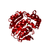
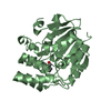
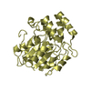
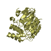
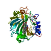
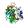
| Entry | Database: PDB / ID: 1a8s | ||||||
|---|---|---|---|---|---|---|---|
| Title | CHLOROPEROXIDASE F/PROPIONATE COMPLEX | ||||||
 Components Components | CHLOROPEROXIDASE F | ||||||
 Keywords Keywords | HALOPEROXIDASE / OXIDOREDUCTASE / PROPIONATE COMPLEX | ||||||
| Function / homology |  Function and homology information Function and homology informationOxidoreductases; Acting on a peroxide as acceptor; Peroxidases / peroxidase activity Similarity search - Function | ||||||
| Biological species |  Pseudomonas fluorescens (bacteria) Pseudomonas fluorescens (bacteria) | ||||||
| Method |  X-RAY DIFFRACTION / SYNCHROTRON / MOLECULAR REPLACEMENT / Resolution: 1.8 Å X-RAY DIFFRACTION / SYNCHROTRON / MOLECULAR REPLACEMENT / Resolution: 1.8 Å | ||||||
 Authors Authors | Hofmann, B. / Toelzer, S. / Pelletier, I. / Altenbuchner, J. / Van Pee, K.-H. / Hecht, H.-J. | ||||||
 Citation Citation | Journal: J.Mol.Biol. / Year: 1998 Title: Structural investigation of the cofactor-free chloroperoxidases. Authors: Hofmann, B. / Tolzer, S. / Pelletier, I. / Altenbuchner, J. / van Pee, K.H. / Hecht, H.J. | ||||||
| History |
|
- Structure visualization
| Structure viewer | Molecule: MolmilJmol/JSmol |
|---|
- Downloads & links
-Download
| PDBx/mmCIF format | 1a8s.cif.gz | 73 KB | Display | PDBx/mmCIF format |
|---|---|---|---|---|
| PDB format | pdb1a8s.ent.gz | 53.7 KB | Display | PDB format |
| PDBx/mmJSON format | 1a8s.json.gz | Tree view | PDBx/mmJSON format | |
| Others |  Other downloads Other downloads |
-Validation report
| Arichive directory | https://data.pdbj.org/pub/pdb/validation_reports/a8/1a8sftp://data.pdbj.org/pub/pdb/validation_reports/a8/1a8s | HTTPS FTP |
|---|
-Related structure data


-Links
 PDBj
PDBj


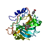
- Assembly
| Deposited unit | 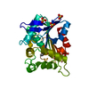
| ||||||||
|---|---|---|---|---|---|---|---|---|---|
| 1 |
| ||||||||
| 2 | x 12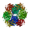
| ||||||||
| 3 | 
| ||||||||
| Unit cell |
|
-Components
| #1: Protein | Mass: 29553.131 Da / Num. of mol.: 1 Source method: isolated from a genetically manipulated source Source: (gene. exp.) Pseudomonas fluorescens (bacteria) / Strain: BL914 / Gene: CPOF / Plasmid: PSK12 / Gene (production host): CPOF / Production host: | ||||
|---|---|---|---|---|---|
| #2: Chemical | ChemComp-SO4 /   Mass: 96.063 Da / Num. of mol.: 4 / Source method: obtained synthetically / Formula: SO4 Mass: 96.063 Da / Num. of mol.: 4 / Source method: obtained synthetically / Formula: SO4#3: Chemical | ChemComp-PPI / | 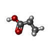  Mass: 74.079 Da / Num. of mol.: 1 / Source method: obtained synthetically / Formula: C3H6O2 Mass: 74.079 Da / Num. of mol.: 1 / Source method: obtained synthetically / Formula: C3H6O2#4: Water | ChemComp-HOH / |  Mass: 18.015 Da / Num. of mol.: 335 / Source method: isolated from a natural source / Formula: H2O Mass: 18.015 Da / Num. of mol.: 335 / Source method: isolated from a natural source / Formula: H2O |
-Experimental details
-Experiment
| Experiment | Method: X-RAY DIFFRACTION / Number of used crystals: 1 |
|---|
- Sample preparation
| Crystal | Density Matthews: 3.36 Å3/Da / Density % sol: 63.1 % | |||||||||||||||
|---|---|---|---|---|---|---|---|---|---|---|---|---|---|---|---|---|
| Crystal grow | pH: 6.6 Details: 1.0 M AMMONIUM SULFATE 50MM CITRATE/PHOSPHATE BUFFER PH 6.6 | |||||||||||||||
| Crystal grow | *PLUS Temperature: 25 ℃ / Method: vapor diffusion, sitting drop | |||||||||||||||
| Components of the solutions | *PLUS
|
-Data collection
| Diffraction | Mean temperature: 90 K |
|---|---|
| Diffraction source | Source: SYNCHROTRON / Site: MPG/DESY, HAMBURG  / Beamline: BW6 / Wavelength: 1.1 / Beamline: BW6 / Wavelength: 1.1 |
| Detector | Type: MARRESEARCH / Detector: IMAGE PLATE / Date: Oct 1, 1997 / Details: MIRROR |
| Radiation | Monochromator: SI(111) / Monochromatic (M) / Laue (L): M / Scattering type: x-ray |
| Radiation wavelength | Wavelength: 1.1 Å / Relative weight: 1 |
| Reflection | Resolution: 1.8→35.5 Å / Num. obs: 33363 / % possible obs: 88.9 % / Observed criterion σ(I): 0 / Redundancy: 2 % / Biso Wilson estimate: 18.38 Å2 / Rmerge(I) obs: 0.069 / Net I/σ(I): 13.1 |
| Reflection shell | Resolution: 1.8→1.88 Å / Redundancy: 1.7 % / Rmerge(I) obs: 0.197 / Mean I/σ(I) obs: 4.9 / % possible all: 80.7 |
| Reflection shell | *PLUS % possible obs: 80.7 % |
- Processing
| Software |
| ||||||||||||||||||||||||||||||||||||||||||||||||||||||||||||||||||||||||||||||||||||
|---|---|---|---|---|---|---|---|---|---|---|---|---|---|---|---|---|---|---|---|---|---|---|---|---|---|---|---|---|---|---|---|---|---|---|---|---|---|---|---|---|---|---|---|---|---|---|---|---|---|---|---|---|---|---|---|---|---|---|---|---|---|---|---|---|---|---|---|---|---|---|---|---|---|---|---|---|---|---|---|---|---|---|---|---|---|
| Refinement | Method to determine structure: MOLECULAR REPLACEMENT Starting model: REFINED COORDINATES OF CHLOROPEROXIDASE L Resolution: 1.8→70 Å / Cross valid method: THROUGHOUT / σ(F): 0
| ||||||||||||||||||||||||||||||||||||||||||||||||||||||||||||||||||||||||||||||||||||
| Displacement parameters | Biso mean: 20.57 Å2 | ||||||||||||||||||||||||||||||||||||||||||||||||||||||||||||||||||||||||||||||||||||
| Refine analyze | Luzzati d res low obs: 8.4 Å / Luzzati sigma a obs: 0.15 Å | ||||||||||||||||||||||||||||||||||||||||||||||||||||||||||||||||||||||||||||||||||||
| Refinement step | Cycle: LAST / Resolution: 1.8→70 Å
| ||||||||||||||||||||||||||||||||||||||||||||||||||||||||||||||||||||||||||||||||||||
| Refine LS restraints |
|
