Protocol: SINGLE WAVELENGTH / Monochromatic (M) / Laue (L): M / Scattering type: x-ray
Radiation wavelength
Wavelength: 0.9537 Å / Relative weight: 1
Reflection
Resolution: 1.8→66.71 Å / Num. obs: 31205 / % possible obs: 93.8 % / Redundancy: 4.1 % / Net I/σ(I): 14.7
-
Processing
Software
Name
Version
Classification
REFMAC
5.8.0158
refinement
iMOSFLM
datareduction
Aimless
datascaling
PHASER
phasing
Refinement
Resolution: 1.8→66.71 Å / Cor.coef. Fo:Fc: 0.969 / Cor.coef. Fo:Fc free: 0.952 / SU B: 2.101 / SU ML: 0.063 / Cross valid method: THROUGHOUT / ESU R: 0.095 / ESU R Free: 0.098 / Details: HYDROGENS HAVE BEEN USED IF PRESENT IN THE INPUT
Rfactor
Num. reflection
% reflection
Selection details
Rfree
0.189
1654
5 %
RANDOM
Rwork
0.152
-
-
-
obs
0.154
31205
93.8 %
-
Solvent computation
Ion probe radii: 0.8 Å / Shrinkage radii: 0.8 Å / VDW probe radii: 1.2 Å
 Movie
Movie Controller

 Yorodumi
 Open data
 Basic information
 Components
 Keywords
 Function and homology information
 X-RAY DIFFRACTION /
  Authors
 Australia, 1items
  Citation
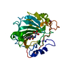
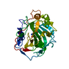
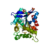
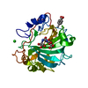
 Structure visualization
 Downloads & links
 Other downloads
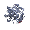
 PDBj
PDBj
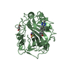
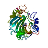
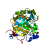
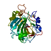
 Assembly


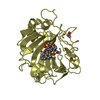
 Mass: 92.094 Da / Num. of mol.: 3 / Source method: obtained synthetically / Formula: C3H8O3
 Mass: 18.015 Da / Num. of mol.: 230 / Source method: isolated from a natural source / Formula: H2O
 Sample preparation
 Processing