 Movie
Movie Controller

[English] 日本語
 Yorodumi
- PDB-1erz: CRYSTAL STRUCTURE OF N-CARBAMYL-D-AMINO ACID AMIDOHYDROLASE WITH ... -
+ Open data

- Basic information
| Entry | Database: PDB / ID: 1erz | ||||||
|---|---|---|---|---|---|---|---|
| Title | CRYSTAL STRUCTURE OF N-CARBAMYL-D-AMINO ACID AMIDOHYDROLASE WITH A NOVEL CATALYTIC FRAMEWORK COMMON TO AMIDOHYDROLASES | ||||||
 Components Components | N-CARBAMYL-D-AMINO ACID AMIDOHYDROLASE | ||||||
 Keywords Keywords | HYDROLASE / four-layer sandwich | ||||||
| Function / homology |  Function and homology information Function and homology informationN-carbamoyl-D-amino-acid hydrolase / N-carbamoyl-D-amino acid hydrolase activity Similarity search - Function | ||||||
| Biological species |  Agrobacterium sp. (bacteria) Agrobacterium sp. (bacteria) | ||||||
| Method |  X-RAY DIFFRACTION / SYNCHROTRON / Resolution: 1.7 Å X-RAY DIFFRACTION / SYNCHROTRON / Resolution: 1.7 Å | ||||||
 Authors Authors | Nakai, T. / Hasegawa, T. / Yamashita, E. / Yamamoto, M. / Kumasaka, T. / Ueki, T. / Nanba, H. / Ikenaka, Y. / Takahashi, S. / Sato, M. / Tsukihara, T. | ||||||
 Citation Citation | Journal: Structure Fold.Des. / Year: 2000 Title: Crystal structure of N-carbamyl-D-amino acid amidohydrolase with a novel catalytic framework common to amidohydrolases. Authors: Nakai, T. / Hasegawa, T. / Yamashita, E. / Yamamoto, M. / Kumasaka, T. / Ueki, T. / Nanba, H. / Ikenaka, Y. / Takahashi, S. / Sato, M. / Tsukihara, T. #1: Journal: BIOSCI.BIOTECHNOL.BIOCHEM. / Year: 1998Title: Isolation of Agrobacterium sp. Strain KNK712 that Produces N-carbamyl-D-amino acid Amidohydrolase, Cloning of the Gene for this Enzyme, and Properties of the Enzyme. Authors: Nanba, H. / Ikenaka, Y. / Yamada, Y. / Yajima, K. / Takano, M. / Takahashi, S. #2: Journal: PATENT / Year: 1999Title: Crystals and Three-dimensional Structure of DCase, and their Use Authors: Nakai, T. / Morikawa, S. / Ishii, K. / Nanba, H. / Yajima, K. / Ikenaka, Y. / Takahashi, S. | ||||||
| History |
|
- Structure visualization
| Structure viewer | Molecule: MolmilJmol/JSmol |
|---|
- Downloads & links
-Download
| PDBx/mmCIF format | 1erz.cif.gz | 144.9 KB | Display | PDBx/mmCIF format |
|---|---|---|---|---|
| PDB format | pdb1erz.ent.gz | 114.9 KB | Display | PDB format |
| PDBx/mmJSON format | 1erz.json.gz | Tree view | PDBx/mmJSON format | |
| Others |  Other downloads Other downloads |
-Validation report
| Arichive directory | https://data.pdbj.org/pub/pdb/validation_reports/er/1erzftp://data.pdbj.org/pub/pdb/validation_reports/er/1erz | HTTPS FTP |
|---|
-Related structure data
| Similar structure data |
|---|


-Links
 PDBj
PDBj- Assembly
| Deposited unit | 
| |||||||||
|---|---|---|---|---|---|---|---|---|---|---|
| 1 | 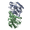
| |||||||||
| Unit cell |
| |||||||||
| Components on special symmetry positions |
| |||||||||
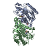
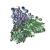
| Details | The biological assembly is a tetramer constructed from chain A and B. A symmetry partner generated by the two-fold. |
-Components
| #1: Protein | Mass: 34199.094 Da / Num. of mol.: 2 Source method: isolated from a genetically manipulated source Source: (gene. exp.) Agrobacterium sp. (bacteria) / Strain: KNK712 / Plasmid: PAD108 / Production host: References: UniProt: P60327, N-carbamoyl-D-amino-acid hydrolase #2: Water | ChemComp-HOH / |  Mass: 18.015 Da / Num. of mol.: 759 / Source method: isolated from a natural source / Formula: H2O Mass: 18.015 Da / Num. of mol.: 759 / Source method: isolated from a natural source / Formula: H2O |
|---|
-Experimental details
-Experiment
| Experiment | Method: X-RAY DIFFRACTION / Number of used crystals: 1 |
|---|
- Sample preparation
| Crystal | Density Matthews: 2.34 Å3/Da / Density % sol: 47.35 % | ||||||||||||||||||||||||
|---|---|---|---|---|---|---|---|---|---|---|---|---|---|---|---|---|---|---|---|---|---|---|---|---|---|
| Crystal grow | Temperature: 293 K / Method: vapor diffusion, sitting drop / pH: 7.5 Details: PEG 6000, lithium chloride, HEPES, pH 7.5, VAPOR DIFFUSION, SITTING DROP, temperature 293K | ||||||||||||||||||||||||
| Crystal grow | *PLUS Temperature: 20 ℃ | ||||||||||||||||||||||||
| Components of the solutions | *PLUS
|
-Data collection
| Diffraction | Mean temperature: 100 K |
|---|---|
| Diffraction source | Source: SYNCHROTRON / Site: SPring-8  / Beamline: BL45XU / Wavelength: 0.9341 / Beamline: BL45XU / Wavelength: 0.9341 |
| Detector | Type: RIGAKU RAXIS IV / Detector: IMAGE PLATE / Date: Oct 13, 1998 |
| Radiation | Protocol: SINGLE WAVELENGTH / Monochromatic (M) / Laue (L): M / Scattering type: x-ray |
| Radiation wavelength | Wavelength: 0.9341 Å / Relative weight: 1 |
| Reflection | Resolution: 1.7→69 Å / Num. all: 68606 / Num. obs: 68596 / % possible obs: 96.1 % / Observed criterion σ(F): 0 / Observed criterion σ(I): 0 / Redundancy: 2.8 % / Biso Wilson estimate: 22.87 Å2 / Rmerge(I) obs: 0.064 / Net I/σ(I): 22.8 |
| Reflection shell | Resolution: 1.7→1.76 Å / Redundancy: 2.3 % / Rmerge(I) obs: 0.207 / Num. unique all: 6418 / % possible all: 91.4 |
| Reflection | *PLUS Num. measured all: 189832 |
| Reflection shell | *PLUS % possible obs: 91.4 % |
- Processing
| Software |
| |||||||||||||||||||||||||
|---|---|---|---|---|---|---|---|---|---|---|---|---|---|---|---|---|---|---|---|---|---|---|---|---|---|---|
| Refinement | Resolution: 1.7→69 Å / σ(F): 0 / σ(I): 0 / Stereochemistry target values: Engh & Huber
| |||||||||||||||||||||||||
| Refinement step | Cycle: LAST / Resolution: 1.7→69 Å
| |||||||||||||||||||||||||
| Refine LS restraints |
|
