 Movie
Movie Controller

[English] 日本語
 Yorodumi
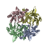
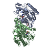
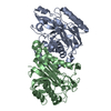
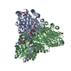
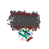
- PDB-1fo6: CRYSTAL STRUCTURE ANALYSIS OF N-CARBAMoYL-D-AMINO-ACID AMIDOHYDROLASE -
+ Open data

- Basic information
| Entry | Database: PDB / ID: 1fo6 | ||||||
|---|---|---|---|---|---|---|---|
| Title | CRYSTAL STRUCTURE ANALYSIS OF N-CARBAMoYL-D-AMINO-ACID AMIDOHYDROLASE | ||||||
 Components Components | N-CARBAMoYL-D-AMINO-ACID AMIDOHYDROLASE | ||||||
 Keywords Keywords | HYDROLASE / four layer a/b fold | ||||||
| Function / homology |  Function and homology information Function and homology informationN-carbamoyl-D-amino-acid hydrolase / N-carbamoyl-D-amino acid hydrolase activity Similarity search - Function | ||||||
| Biological species |  Agrobacterium tumefaciens (bacteria) Agrobacterium tumefaciens (bacteria) | ||||||
| Method |  X-RAY DIFFRACTION / SYNCHROTRON / Resolution: 1.95 Å X-RAY DIFFRACTION / SYNCHROTRON / Resolution: 1.95 Å | ||||||
 Authors Authors | Wang, W.-C. / Hsu, W.-H. / Chien, F.-T. / Chen, C.-Y. | ||||||
 Citation Citation | Journal: J.Mol.Biol. / Year: 2001 Title: Crystal structure and site-directed mutagenesis studies of N-carbamoyl-D-amino-acid amidohydrolase from Agrobacterium radiobacter reveals a homotetramer and insight into a catalytic cleft. Authors: Wang, W.C. / Hsu, W.H. / Chien, F.T. / Chen, C.Y. #1: Journal: Acta Crystallogr.,Sect.D / Year: 1999Title: Expression, crystallization and preliminary X-ray diffraction studies of N-carbamyl-D-amino-acid amidohydrolase from Agrobacterium radiobacter Authors: Hsu, W.-H. / Chien, F.-T. / Hsu, C.L. / Wang, T.C. / Yuan, H.S. / Wang, W.-C. | ||||||
| History |
|
- Structure visualization
| Structure viewer | Molecule: MolmilJmol/JSmol |
|---|
- Downloads & links
-Download
| PDBx/mmCIF format | 1fo6.cif.gz | 257.6 KB | Display | PDBx/mmCIF format |
|---|---|---|---|---|
| PDB format | pdb1fo6.ent.gz | 206.7 KB | Display | PDB format |
| PDBx/mmJSON format | 1fo6.json.gz | Tree view | PDBx/mmJSON format | |
| Others |  Other downloads Other downloads |
-Validation report
| Arichive directory | https://data.pdbj.org/pub/pdb/validation_reports/fo/1fo6ftp://data.pdbj.org/pub/pdb/validation_reports/fo/1fo6 | HTTPS FTP |
|---|
-Related structure data
| Similar structure data |
|---|


-Links
 PDBj
PDBj
- Assembly
| Deposited unit | 
| ||||||||
|---|---|---|---|---|---|---|---|---|---|
| 1 |
| ||||||||
| Unit cell |
|
-Components
| #1: Protein | Mass: 34209.062 Da / Num. of mol.: 4 Source method: isolated from a genetically manipulated source Source: (gene. exp.) Agrobacterium tumefaciens (bacteria) / Plasmid: PQE30 / Production host: #2: Chemical |   Mass: 131.293 Da / Num. of mol.: 2 / Source method: obtained synthetically / Formula: Xe Mass: 131.293 Da / Num. of mol.: 2 / Source method: obtained synthetically / Formula: Xe#3: Water | ChemComp-HOH / |  Mass: 18.015 Da / Num. of mol.: 722 / Source method: isolated from a natural source / Formula: H2O Mass: 18.015 Da / Num. of mol.: 722 / Source method: isolated from a natural source / Formula: H2O |
|---|
-Experimental details
-Experiment
| Experiment | Method: X-RAY DIFFRACTION / Number of used crystals: 6 |
|---|
- Sample preparation
| Crystal | Density Matthews: 2.37 Å3/Da / Density % sol: 48.05 % | |||||||||||||||||||||||||||||||||||
|---|---|---|---|---|---|---|---|---|---|---|---|---|---|---|---|---|---|---|---|---|---|---|---|---|---|---|---|---|---|---|---|---|---|---|---|---|
| Crystal grow | Temperature: 296.5 K / Method: vapor diffusion, hanging drop / pH: 7 Details: Lithium sulfate, HEPES , pH 7.0, VAPOR DIFFUSION, HANGING DROP, temperature 296.5K | |||||||||||||||||||||||||||||||||||
| Crystal grow | *PLUS Temperature: 23 ℃ / pH: 7.5 | |||||||||||||||||||||||||||||||||||
| Components of the solutions | *PLUS
|
-Data collection
| Diffraction |
| ||||||||||||||||||
|---|---|---|---|---|---|---|---|---|---|---|---|---|---|---|---|---|---|---|---|
| Diffraction source |
| ||||||||||||||||||
| Detector |
| ||||||||||||||||||
| Radiation | Protocol: SINGLE WAVELENGTH / Monochromatic (M) / Laue (L): M / Scattering type: x-ray | ||||||||||||||||||
| Radiation wavelength |
| ||||||||||||||||||
| Reflection | Resolution: 1.95→30 Å / Num. all: 93420 / Num. obs: 93415 / % possible obs: 99.9 % / Redundancy: 7.4 % / Rmerge(I) obs: 0.073 / Net I/σ(I): 7.5 | ||||||||||||||||||
| Reflection shell | Resolution: 1.95→2 Å / Redundancy: 7.4 % / Rmerge(I) obs: 0.459 / Num. unique all: 6865 / % possible all: 99.9 | ||||||||||||||||||
| Reflection | *PLUS Lowest resolution: 30 Å | ||||||||||||||||||
| Reflection shell | *PLUS % possible obs: 99.8 % |

- Processing
| Software |
| ||||||||||||||||||||
|---|---|---|---|---|---|---|---|---|---|---|---|---|---|---|---|---|---|---|---|---|---|
| Refinement | Resolution: 1.95→30 Å / σ(F): 0 / σ(I): 0 Details: Refinement of Macromolecular Structures by the Maximum-Likelihood Method
| ||||||||||||||||||||
| Refinement step | Cycle: LAST / Resolution: 1.95→30 Å
| ||||||||||||||||||||
| Refine LS restraints |
| ||||||||||||||||||||
| Software | *PLUS Name: REFMAC / Classification: refinement | ||||||||||||||||||||
| Refinement | *PLUS Lowest resolution: 30 Å / σ(F): 0 / % reflection Rfree: 5 % / Rfactor Rwork: 0.187 | ||||||||||||||||||||
| Solvent computation | *PLUS | ||||||||||||||||||||
| Displacement parameters | *PLUS | ||||||||||||||||||||
| Refine LS restraints | *PLUS
|
