 Movie
Movie Controller

[English] 日本語
 Yorodumi
- PDB-2ggl: The mutant A222C of Agrobacterium radiobacter N-carbamoyl-D-amino... -
+ Open data

- Basic information
| Entry | Database: PDB / ID: 2ggl | |||||||||
|---|---|---|---|---|---|---|---|---|---|---|
| Title | The mutant A222C of Agrobacterium radiobacter N-carbamoyl-D-amino acid amidohydrolase | |||||||||
 Components Components | N-carbamoyl-D-amino acid amidohydrolase | |||||||||
 Keywords Keywords | HYDROLASE / N-Carbamoyl-D-Amino-Acid Amidohydrolase | |||||||||
| Function / homology |  Function and homology information Function and homology informationN-carbamoyl-D-amino-acid hydrolase / N-carbamoyl-D-amino acid hydrolase activity Similarity search - Function | |||||||||
| Biological species |  Agrobacterium tumefaciens (bacteria) Agrobacterium tumefaciens (bacteria) | |||||||||
| Method |  X-RAY DIFFRACTION / MOLECULAR REPLACEMENT / Resolution: 2.4 Å X-RAY DIFFRACTION / MOLECULAR REPLACEMENT / Resolution: 2.4 Å | |||||||||
 Authors Authors | Wang, W.C. / Chiu, W.C. / You, J.Y. | |||||||||
 Citation Citation | Journal: J.Mol.Biol. / Year: 2006 Title: Structure-Stability-Activity Relationship in Covalently Cross-linked N-Carbamoyl d-Amino acid Amidohydrolase and N-Acylamino acid Racemase. Authors: Chiu, W.C. / You, J.Y. / Liu, J.S. / Hsu, S.K. / Hsu, W.H. / Shih, C.H. / Hwang, J.K. / Wang, W.C. | |||||||||
| History |
|
- Structure visualization
| Structure viewer | Molecule: MolmilJmol/JSmol |
|---|
- Downloads & links
-Download
| PDBx/mmCIF format | 2ggl.cif.gz | 247.5 KB | Display | PDBx/mmCIF format |
|---|---|---|---|---|
| PDB format | pdb2ggl.ent.gz | 201.2 KB | Display | PDB format |
| PDBx/mmJSON format | 2ggl.json.gz | Tree view | PDBx/mmJSON format | |
| Others |  Other downloads Other downloads |
-Validation report
| Arichive directory | https://data.pdbj.org/pub/pdb/validation_reports/gg/2gglftp://data.pdbj.org/pub/pdb/validation_reports/gg/2ggl | HTTPS FTP |
|---|
-Related structure data
| Related structure data |  2gggC 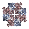 2gghC  2ggiC  2ggjC 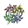 2ggkC 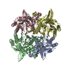 1fo6S S: Starting model for refinement C: citing same article ( |
|---|---|
| Similar structure data |


-Links
 PDBj
PDBj- Assembly
| Deposited unit | 
| ||||||||
|---|---|---|---|---|---|---|---|---|---|
| 1 |
| ||||||||
| 2 | 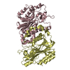
| ||||||||
| 3 | 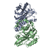
| ||||||||
| Unit cell |
| ||||||||
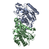
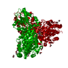
| Details | The crystal structure of D-NCAase A222C mutant reveals a tetramer with 222 symmetry. |
-Components
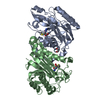
| #1: Protein | Mass: 34241.129 Da / Num. of mol.: 4 / Mutation: A222C Source method: isolated from a genetically manipulated source Source: (gene. exp.) Agrobacterium tumefaciens (bacteria) / Plasmid: pQE30 / Production host: References: UniProt: Q44185, N-carbamoyl-D-amino-acid hydrolase #2: Water | ChemComp-HOH / |  Mass: 18.015 Da / Num. of mol.: 402 / Source method: isolated from a natural source / Formula: H2O Mass: 18.015 Da / Num. of mol.: 402 / Source method: isolated from a natural source / Formula: H2O |
|---|
-Experimental details
-Experiment
| Experiment | Method: X-RAY DIFFRACTION / Number of used crystals: 1 |
|---|
- Sample preparation
| Crystal | Density Matthews: 2.37 Å3/Da / Density % sol: 48.08 % |
|---|---|
| Crystal grow | Temperature: 298 K / Method: vapor diffusion, hanging drop / pH: 7 Details: lithium sulfate, HEPES buffer, pH 7.0, VAPOR DIFFUSION, HANGING DROP, temperature 298K |
-Data collection
| Diffraction | Mean temperature: 113 K |
|---|---|
| Diffraction source | Source: ROTATING ANODE / Type: RIGAKU RU300 / Wavelength: 1.5418 Å |
| Detector | Type: RIGAKU RAXIS IV / Detector: IMAGE PLATE / Date: Apr 13, 2003 |
| Radiation | Protocol: SINGLE WAVELENGTH / Monochromatic (M) / Laue (L): M / Scattering type: x-ray |
| Radiation wavelength | Wavelength: 1.5418 Å / Relative weight: 1 |
| Reflection | Resolution: 2.4→30 Å / Num. obs: 50489 / % possible obs: 94.8 % |
| Reflection shell | Resolution: 2.4→2.49 Å / % possible all: 93.6 |
- Processing
| Software |
| ||||||||||||||||||||||||||||||||||||||||||||||||||||||||||||||||||||||
|---|---|---|---|---|---|---|---|---|---|---|---|---|---|---|---|---|---|---|---|---|---|---|---|---|---|---|---|---|---|---|---|---|---|---|---|---|---|---|---|---|---|---|---|---|---|---|---|---|---|---|---|---|---|---|---|---|---|---|---|---|---|---|---|---|---|---|---|---|---|---|---|
| Refinement | Method to determine structure: MOLECULAR REPLACEMENT Starting model: 1FO6 Resolution: 2.4→30 Å / Cor.coef. Fo:Fc: 0.959 / Cor.coef. Fo:Fc free: 0.938 / SU B: 6.977 / SU ML: 0.162 / Cross valid method: THROUGHOUT / ESU R: 0.335 / ESU R Free: 0.243 / Stereochemistry target values: MAXIMUM LIKELIHOOD / Details: HYDROGENS HAVE BEEN ADDED IN THE RIDING POSITIONS
| ||||||||||||||||||||||||||||||||||||||||||||||||||||||||||||||||||||||
| Solvent computation | Ion probe radii: 0.8 Å / Shrinkage radii: 0.8 Å / VDW probe radii: 1.2 Å / Solvent model: MASK | ||||||||||||||||||||||||||||||||||||||||||||||||||||||||||||||||||||||
| Displacement parameters | Biso mean: 32.097 Å2
| ||||||||||||||||||||||||||||||||||||||||||||||||||||||||||||||||||||||
| Refinement step | Cycle: LAST / Resolution: 2.4→30 Å
| ||||||||||||||||||||||||||||||||||||||||||||||||||||||||||||||||||||||
| Refine LS restraints |
| ||||||||||||||||||||||||||||||||||||||||||||||||||||||||||||||||||||||
| LS refinement shell | Resolution: 2.4→2.462 Å / Total num. of bins used: 20
|
