 Movie
Movie Controller

[English] 日本語
 Yorodumi
- PDB-1ee6: CRYSTAL STRUCTURE OF PECTATE LYASE FROM BACILLUS SP. STRAIN KSM-P15. -
+ Open data

- Basic information
| Entry | Database: PDB / ID: 1ee6 | ||||||
|---|---|---|---|---|---|---|---|
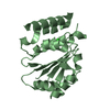
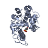
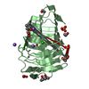
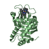
| Title | CRYSTAL STRUCTURE OF PECTATE LYASE FROM BACILLUS SP. STRAIN KSM-P15. | ||||||
 Components Components | PECTATE LYASE | ||||||
 Keywords Keywords | LYASE / Parallel beta-helix / High-alkaline / Low-Molecular-Weight | ||||||
| Function / homology |  Function and homology information Function and homology informationpectin biosynthetic process / polygalacturonase activity / pectate lyase / pectate lyase activity / calcium ion binding / extracellular region Similarity search - Function | ||||||
| Biological species |  | ||||||
| Method |  X-RAY DIFFRACTION / Resolution: 2.3 Å X-RAY DIFFRACTION / Resolution: 2.3 Å | ||||||
 Authors Authors | Akita, M. / Suzuki, A. / Kobayashi, T. / Ito, S. / Yamane, T. | ||||||
 Citation Citation | Journal: Acta Crystallogr.,Sect.D / Year: 2001 Title: The first structure of pectate lyase belonging to polysaccharide lyase family 3. Authors: Akita, M. / Suzuki, A. / Kobayashi, T. / Ito, S. / Yamane, T. | ||||||
| History |
|
- Structure visualization
| Structure viewer | Molecule: MolmilJmol/JSmol |
|---|
- Downloads & links
-Download
| PDBx/mmCIF format | 1ee6.cif.gz | 53 KB | Display | PDBx/mmCIF format |
|---|---|---|---|---|
| PDB format | pdb1ee6.ent.gz | 37.1 KB | Display | PDB format |
| PDBx/mmJSON format | 1ee6.json.gz | Tree view | PDBx/mmJSON format | |
| Others |  Other downloads Other downloads |
-Validation report
| Arichive directory | https://data.pdbj.org/pub/pdb/validation_reports/ee/1ee6ftp://data.pdbj.org/pub/pdb/validation_reports/ee/1ee6 | HTTPS FTP |
|---|
-Related structure data
| Similar structure data |
|---|


-Links
 PDBj
PDBj- Assembly
| Deposited unit | 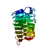
| ||||||||
|---|---|---|---|---|---|---|---|---|---|
| 1 |
| ||||||||
| Unit cell |
|
-Components
| #1: Protein | Mass: 20947.287 Da / Num. of mol.: 1 / Source method: isolated from a natural source / Source: (natural) |
|---|---|
| #2: Chemical | ChemComp-CA /   Mass: 40.078 Da / Num. of mol.: 1 / Source method: obtained synthetically / Formula: Ca Mass: 40.078 Da / Num. of mol.: 1 / Source method: obtained synthetically / Formula: Ca |
| #3: Water | ChemComp-HOH /  Mass: 18.015 Da / Num. of mol.: 170 / Source method: isolated from a natural source / Formula: H2O Mass: 18.015 Da / Num. of mol.: 170 / Source method: isolated from a natural source / Formula: H2O |
| Has protein modification | Y |
-Experimental details
-Experiment
| Experiment | Method: X-RAY DIFFRACTION / Number of used crystals: 1 |
|---|
- Sample preparation
| Crystal | Density Matthews: 2.57 Å3/Da / Density % sol: 52.14 % | ||||||||||||||||||||||||||||||||||||
|---|---|---|---|---|---|---|---|---|---|---|---|---|---|---|---|---|---|---|---|---|---|---|---|---|---|---|---|---|---|---|---|---|---|---|---|---|---|
| Crystal grow | Temperature: 277 K / Method: vapor diffusion, hanging drop / pH: 6.7 Details: PEG 8000, pH 6.7, VAPOR DIFFUSION, HANGING DROP, temperature 277.0K | ||||||||||||||||||||||||||||||||||||
| Crystal grow | *PLUS Temperature: 277 K / pH: 7.5 | ||||||||||||||||||||||||||||||||||||
| Components of the solutions | *PLUS
|
-Data collection
| Diffraction | Mean temperature: 100 K |
|---|---|
| Diffraction source | Source: ROTATING ANODE / Type: RIGAKU RU300 / Wavelength: 1.5418 |
| Detector | Type: RIGAKU RAXIS IV++ / Detector: IMAGE PLATE / Date: Jan 1, 1999 |
| Radiation | Protocol: SINGLE WAVELENGTH / Monochromatic (M) / Laue (L): M / Scattering type: x-ray |
| Radiation wavelength | Wavelength: 1.5418 Å / Relative weight: 1 |
| Reflection | Resolution: 2.3→20 Å / Num. obs: 10063 / % possible obs: 99.3 % / Observed criterion σ(F): 0 / Observed criterion σ(I): 0 / Redundancy: 3.8 % / Rmerge(I) obs: 0.069 / Net I/σ(I): 12 |
| Reflection shell | Resolution: 2.3→2.38 Å / Rmerge(I) obs: 0.204 / % possible all: 98.8 |
| Reflection shell | *PLUS % possible obs: 98.8 % |
- Processing
| Software |
| ||||||||||||||||||||
|---|---|---|---|---|---|---|---|---|---|---|---|---|---|---|---|---|---|---|---|---|---|
| Refinement | Resolution: 2.3→10 Å / σ(F): 3 / σ(I): 0 Details: Four disordered side-chain(1A,7E,20K,39K) were excluded in refinement stage.
| ||||||||||||||||||||
| Refinement step | Cycle: LAST / Resolution: 2.3→10 Å
| ||||||||||||||||||||
| Refine LS restraints |
| ||||||||||||||||||||
| Software | *PLUS Name: X-PLOR / Version: 3.1 / Classification: refinement | ||||||||||||||||||||
| Refinement | *PLUS Highest resolution: 2.3 Å / Lowest resolution: 10 Å / σ(F): 3 | ||||||||||||||||||||
| Solvent computation | *PLUS | ||||||||||||||||||||
| Displacement parameters | *PLUS | ||||||||||||||||||||
| Refine LS restraints | *PLUS Type: x_angle_deg / Dev ideal: 2.7 |
