 Movie
Movie Controller

[English] 日本語
 Yorodumi
- PDB-3t9g: The crystal structure of family 3 pectate lyase from Caldicellulo... -
+ Open data

- Basic information
| Entry | Database: PDB / ID: 3t9g | ||||||
|---|---|---|---|---|---|---|---|
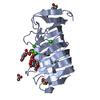
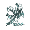
| Title | The crystal structure of family 3 pectate lyase from Caldicellulosiruptor bescii | ||||||
 Components Components | Pectate lyase | ||||||
 Keywords Keywords | LYASE / PL3 / parallel beta-helix / pectate lyase | ||||||
| Function / homology |  Function and homology information Function and homology informationpectate lyase / pectate lyase activity / hydrolase activity / extracellular region / metal ion binding Similarity search - Function | ||||||
| Biological species |   Caldicellulosiruptor bescii (bacteria) Caldicellulosiruptor bescii (bacteria) | ||||||
| Method |  X-RAY DIFFRACTION / MOLECULAR REPLACEMENT / Resolution: 1.5 Å X-RAY DIFFRACTION / MOLECULAR REPLACEMENT / Resolution: 1.5 Å | ||||||
 Authors Authors | Alahuhta, P.M. / Lunin, V.V. | ||||||
 Citation Citation | Journal: Acta Crystallogr.,Sect.F / Year: 2011 Title: A 1.5 A resolution X-ray structure of the catalytic module of Caldicellulosiruptor bescii family 3 pectate lyase. Authors: Alahuhta, M. / Chandrayan, P. / Kataeva, I. / Adams, M.W. / Himmel, M.E. / Lunin, V.V. | ||||||
| History |
|
- Structure visualization
| Structure viewer | Molecule: MolmilJmol/JSmol |
|---|
- Downloads & links
-Download
| PDBx/mmCIF format | 3t9g.cif.gz | 114.8 KB | Display | PDBx/mmCIF format |
|---|---|---|---|---|
| PDB format | pdb3t9g.ent.gz | 87.6 KB | Display | PDB format |
| PDBx/mmJSON format | 3t9g.json.gz | Tree view | PDBx/mmJSON format | |
| Others |  Other downloads Other downloads |
-Validation report
| Arichive directory | https://data.pdbj.org/pub/pdb/validation_reports/t9/3t9gftp://data.pdbj.org/pub/pdb/validation_reports/t9/3t9g | HTTPS FTP |
|---|
-Related structure data
| Related structure data |  1ee6S S: Starting model for refinement |
|---|---|
| Similar structure data |


-Links
 PDBj
PDBj- Assembly
| Deposited unit | 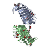
| |||||||||
|---|---|---|---|---|---|---|---|---|---|---|
| 1 | 
| |||||||||
| 2 | 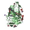
| |||||||||
| 3 | 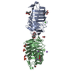
| |||||||||
| Unit cell |
| |||||||||
| Components on special symmetry positions |
|
-Components
-Protein , 1 types, 2 molecules AB
| #1: Protein | Mass: 21132.855 Da / Num. of mol.: 2 / Fragment: unp residues 268-460 Source method: isolated from a genetically manipulated source Source: (gene. exp.) Caldicellulosiruptor bescii (bacteria) / Gene: Athe_1854, Cbes_1854 / Plasmid: pET-45b / Production host: |
|---|
-Non-polymers , 10 types, 551 molecules 


| #2: Chemical |  Mass: 40.078 Da / Num. of mol.: 2 / Source method: obtained synthetically / Formula: Ca Mass: 40.078 Da / Num. of mol.: 2 / Source method: obtained synthetically / Formula: Ca#3: Chemical | ChemComp-MRD / (  Mass: 118.174 Da / Num. of mol.: 4 / Source method: obtained synthetically / Formula: C6H14O2 / Comment: precipitant*YM Mass: 118.174 Da / Num. of mol.: 4 / Source method: obtained synthetically / Formula: C6H14O2 / Comment: precipitant*YM#4: Chemical | ChemComp-MPD / (  Mass: 118.174 Da / Num. of mol.: 4 / Source method: obtained synthetically / Formula: C6H14O2 / Comment: precipitant*YM Mass: 118.174 Da / Num. of mol.: 4 / Source method: obtained synthetically / Formula: C6H14O2 / Comment: precipitant*YM#5: Chemical | ChemComp-NA /  Mass: 22.990 Da / Num. of mol.: 4 / Source method: obtained synthetically / Formula: Na Mass: 22.990 Da / Num. of mol.: 4 / Source method: obtained synthetically / Formula: Na#6: Chemical | ChemComp-GOL /  Mass: 92.094 Da / Num. of mol.: 4 / Source method: obtained synthetically / Formula: C3H8O3 Mass: 92.094 Da / Num. of mol.: 4 / Source method: obtained synthetically / Formula: C3H8O3#7: Chemical | ChemComp-CL / |  Mass: 35.453 Da / Num. of mol.: 1 / Source method: obtained synthetically / Formula: Cl Mass: 35.453 Da / Num. of mol.: 1 / Source method: obtained synthetically / Formula: Cl#8: Chemical |  Mass: 62.068 Da / Num. of mol.: 2 / Source method: obtained synthetically / Formula: C2H6O2 Mass: 62.068 Da / Num. of mol.: 2 / Source method: obtained synthetically / Formula: C2H6O2#9: Chemical |  Mass: 59.044 Da / Num. of mol.: 2 / Source method: obtained synthetically / Formula: C2H3O2 Mass: 59.044 Da / Num. of mol.: 2 / Source method: obtained synthetically / Formula: C2H3O2#10: Chemical |  Mass: 94.971 Da / Num. of mol.: 2 / Source method: obtained synthetically / Formula: PO4 Mass: 94.971 Da / Num. of mol.: 2 / Source method: obtained synthetically / Formula: PO4#11: Water | ChemComp-HOH / | Mass: 18.015 Da / Num. of mol.: 526 / Source method: isolated from a natural source / Formula: H2O |
|---|
-Experimental details
-Experiment
| Experiment | Method: X-RAY DIFFRACTION / Number of used crystals: 1 |
|---|
- Sample preparation
| Crystal | Density Matthews: 2.57 Å3/Da / Density % sol: 52.11 % |
|---|---|
| Crystal grow | Temperature: 293 K / Method: vapor diffusion, sitting drop / pH: 8.5 Details: 0.2 M ammonium phosphate monobasic, 0.1 M Tris, 50% v/v (+/-)-2-methyl-2,4-pentanediol, pH 8.5, VAPOR DIFFUSION, SITTING DROP, temperature 293K |
-Data collection
| Diffraction | Mean temperature: 100 K |
|---|---|
| Diffraction source | Source: ROTATING ANODE / Type: BRUKER AXS MICROSTAR / Wavelength: 1.5418 Å |
| Detector | Type: Bruker Platinum 135 / Detector: CCD / Date: Jul 29, 2011 / Details: HELIOS MIRRORS |
| Radiation | Monochromator: HELIOS MIRRORS / Protocol: SINGLE WAVELENGTH / Monochromatic (M) / Laue (L): M / Scattering type: x-ray |
| Radiation wavelength | Wavelength: 1.5418 Å / Relative weight: 1 |
| Reflection | Resolution: 1.5→25 Å / Num. all: 70692 / Num. obs: 70692 / % possible obs: 99.6 % / Redundancy: 6.15 % / Rsym value: 0.0446 / Net I/σ(I): 24.73 |
| Reflection shell | Resolution: 1.5→1.6 Å / Redundancy: 3.97 % / Mean I/σ(I) obs: 8.23 / Num. unique all: 12550 / Rsym value: 0.1495 / % possible all: 98.5 |
- Processing
| Software |
| |||||||||||||||||||||||||||||||||||||||||||||||||||||||||||||||||||||||||||||||||||||
|---|---|---|---|---|---|---|---|---|---|---|---|---|---|---|---|---|---|---|---|---|---|---|---|---|---|---|---|---|---|---|---|---|---|---|---|---|---|---|---|---|---|---|---|---|---|---|---|---|---|---|---|---|---|---|---|---|---|---|---|---|---|---|---|---|---|---|---|---|---|---|---|---|---|---|---|---|---|---|---|---|---|---|---|---|---|---|
| Refinement | Method to determine structure: MOLECULAR REPLACEMENT Starting model: 1EE6 Resolution: 1.5→25 Å / Cor.coef. Fo:Fc: 0.972 / Cor.coef. Fo:Fc free: 0.961 / SU B: 1.095 / SU ML: 0.041 / Cross valid method: THROUGHOUT / ESU R Free: 0.066 / Stereochemistry target values: MAXIMUM LIKELIHOOD / Details: HYDROGENS HAVE BEEN ADDED IN THE RIDING POSITIONS
| |||||||||||||||||||||||||||||||||||||||||||||||||||||||||||||||||||||||||||||||||||||
| Solvent computation | Ion probe radii: 0.8 Å / Shrinkage radii: 0.8 Å / VDW probe radii: 1.4 Å / Solvent model: MASK | |||||||||||||||||||||||||||||||||||||||||||||||||||||||||||||||||||||||||||||||||||||
| Displacement parameters | Biso mean: 10.039 Å2
| |||||||||||||||||||||||||||||||||||||||||||||||||||||||||||||||||||||||||||||||||||||
| Refinement step | Cycle: LAST / Resolution: 1.5→25 Å
| |||||||||||||||||||||||||||||||||||||||||||||||||||||||||||||||||||||||||||||||||||||
| Refine LS restraints |
| |||||||||||||||||||||||||||||||||||||||||||||||||||||||||||||||||||||||||||||||||||||
| LS refinement shell | Resolution: 1.497→1.536 Å / Total num. of bins used: 20
|
