 ムービー
ムービー コントローラー
 構造ビューア
 EMN検索について


-検索条件
-検索結果
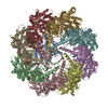
検索 (著者・登録者: sun & dm)の結果123件中、1から50件目までを表示しています
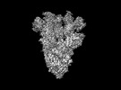
EMDB-28198: 
Cryo-EM map of SARS-CoV-2 Omicron BA.2 spike in complex with LLNL-199

EMDB-28199:
Cryo-EM map of SARS-CoV-2 Omicron BA.2 spike in complex with 2130-1-0114-112
PDB-8ekd:
Cryo-EM map of SARS-CoV-2 Omicron BA.2 spike in complex with 2130-1-0114-112

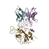
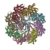
EMDB-35122:
Human TRiC-PhLP2A complex in the closed state

EMDB-35199:
The focused refinement of CCT3-PhLP2A from TRiC-PhLP2A complex in the open state

PDB-8i1u:
Human TRiC-PhLP2A complex in the closed state

PDB-8i6j:
The focused refinement of CCT3-PhLP2A from TRiC-PhLP2A complex in the open state

EMDB-35284:
Human TRiC-PhLP2A complex in the open state

PDB-8i9u:
Human TRiC-PhLP2A complex in the open state

EMDB-35280:
The focused refinement of CCT4-PhLP2A from TRiC-PhLP2A complex in the open state

EMDB-35335:
Human TRiC-PhLP2A-actin complex in the closed state

PDB-8i9q:
The focused refinement of CCT4-PhLP2A from TRiC-PhLP2A complex in the open state

PDB-8ib8:
Human TRiC-PhLP2A-actin complex in the closed state
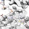
EMDB-29307:
Structure of WT HIV-1 intasome bound to Dolutegravir

EMDB-29309:
Structure of E138K HIV-1 intasome with Dolutegravir bound

EMDB-29312:
Structure of E138K HIV-1 intasome with Dolutegravir bound

EMDB-29313:
Structure of Q148K HIV-1 intasome with Dolutegravir bound

EMDB-29315:
Structure of E138K/G140A HIV-1 intasome with Dolutegravir bound

EMDB-29317:
Structure of E138K/Q148K HIV-1 intasome with Dolutegravir bound

EMDB-29318:
Structure of G140A/Q148K HIV-1 intasome with Dolutegravir bound

EMDB-29319:
Structure of E138K/G140A/Q148K HIV-1 intasome with Dolutegravir bound

EMDB-29320:
Structure of E138K/G140A/Q148R HIV-1 intasome with Dolutegravir bound

EMDB-29321:
Structure of E138K/G140S/Q148H HIV-1 intasome with Dolutegravir bound

EMDB-29322:
Structure of E138K/G140A/Q148K HIV-1 intasome with 4d bound

PDB-8fn7:
Structure of WT HIV-1 intasome bound to Dolutegravir

PDB-8fnd:
Structure of E138K HIV-1 intasome with Dolutegravir bound

PDB-8fng:
Structure of E138K HIV-1 intasome with Dolutegravir bound

PDB-8fnh:
Structure of Q148K HIV-1 intasome with Dolutegravir bound

PDB-8fnj:
Structure of E138K/G140A HIV-1 intasome with Dolutegravir bound

PDB-8fnl:
Structure of E138K/Q148K HIV-1 intasome with Dolutegravir bound

PDB-8fnm:
Structure of G140A/Q148K HIV-1 intasome with Dolutegravir bound

PDB-8fnn:
Structure of E138K/G140A/Q148K HIV-1 intasome with Dolutegravir bound

PDB-8fno:
Structure of E138K/G140A/Q148R HIV-1 intasome with Dolutegravir bound

PDB-8fnp:
Structure of E138K/G140S/Q148H HIV-1 intasome with Dolutegravir bound

PDB-8fnq:
Structure of E138K/G140A/Q148K HIV-1 intasome with 4d bound
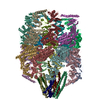
EMDB-28139:
Toxoplasma gondii apical complex (ionophore stimulated)
EMDB-28140:
Toxoplasma gondii apical complex (non-stimulated)
EMDB-32822:
An apo TRiC map

EMDB-26089:
The beta-tubulin folding intermediate I

EMDB-26120:
The beta-tubulin folding intermediate II
EMDB-26123:
The beta-tubulin folding intermediate III

EMDB-26131:
The beta-tubulin folding intermediate IV
PDB-7trg:
The beta-tubulin folding intermediate I

PDB-7ttn:
The beta-tubulin folding intermediate II
PDB-7ttt:
The beta-tubulin folding intermediate III
PDB-7tub:
The beta-tubulin folding intermediate IV

EMDB-32823:
Prefoldin-tubulin-TRiC complex
PDB-7wu7:
Prefoldin-tubulin-TRiC complex
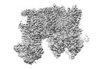
EMDB-26193:
Cryo-EM structure of the pancreatic ATP-sensitive potassium channel bound to ATP and repaglinide with Kir6.2-CTD in the up conformation
EMDB-26194:
Cryo-EM structure of the pancreatic ATP-sensitive potassium channel bound to ATP and repaglinide with Kir6.2-CTD in the down conformation
ページ:
 wwPDBはEMDBデータモデルのバージョン3へ移行します
