Journal: PNAS Nexus / Year: 2022 Title: Cryo-electron tomography with mixed-scale dense neural networks reveals key steps in deployment of invasion machinery. Authors: Li-Av Segev-Zarko / Peter D Dahlberg / Stella Y Sun / Daniël M Pelt / Chi Yong Kim / Elizabeth S Egan / James A Sethian / Wah Chiu / John C Boothroyd / Abstract: Host cell invasion by intracellular, eukaryotic parasites within the phylum Apicomplexa is a remarkable and active process involving the coordinated action of apical organelles and other structures. ...Host cell invasion by intracellular, eukaryotic parasites within the phylum Apicomplexa is a remarkable and active process involving the coordinated action of apical organelles and other structures. To date, capturing how these structures interact during invasion has been difficult to observe in detail. Here, we used cryogenic electron tomography to image the apical complex of tachyzoites under conditions that mimic resting parasites and those primed to invade through stimulation with calcium ionophore. Through the application of mixed-scale dense networks for image processing, we developed a highly efficient pipeline for annotation of tomograms, enabling us to identify and extract densities of relevant subcellular organelles and accurately analyze features in 3-D. The results reveal a dramatic change in the shape of the anteriorly located apical vesicle upon its apparent fusion with a rhoptry that occurs only in the stimulated parasites. We also present information indicating that this vesicle originates from the vesicles that parallel the intraconoidal microtubules and that the latter two structures are linked by a novel tether. We show that a rosette structure previously proposed to be involved in rhoptry secretion is associated with apical vesicles beyond just the most anterior one. This result, suggesting multiple vesicles are primed to enable rhoptry secretion, may shed light on the mechanisms employs to enable repeated invasion attempts. Using the same approach, we examine merozoites and show that they too possess an apical vesicle just beneath a rosette, demonstrating evolutionary conservation of this overall subcellular organization.
pH: 7.2 / Component - Concentration: 1.0 x / Component - Name: Endo buffer Details: 45 mM potassium sulfate, 106 mM sucrose, 10 mM magnesium sulfate, 20 mM Tris buffer pH 7.2, 5 mM glucose and 0.35% bovine serum albumin
Grid
Model: EMS Lacey Carbon / Material: COPPER / Support film - Material: CARBON / Support film - topology: LACEY / Pretreatment - Type: PLASMA CLEANING
Vitrification
Cryogen name: ETHANE / Chamber humidity: 90 % / Chamber temperature: 297 K / Instrument: LEICA EM GP
Details
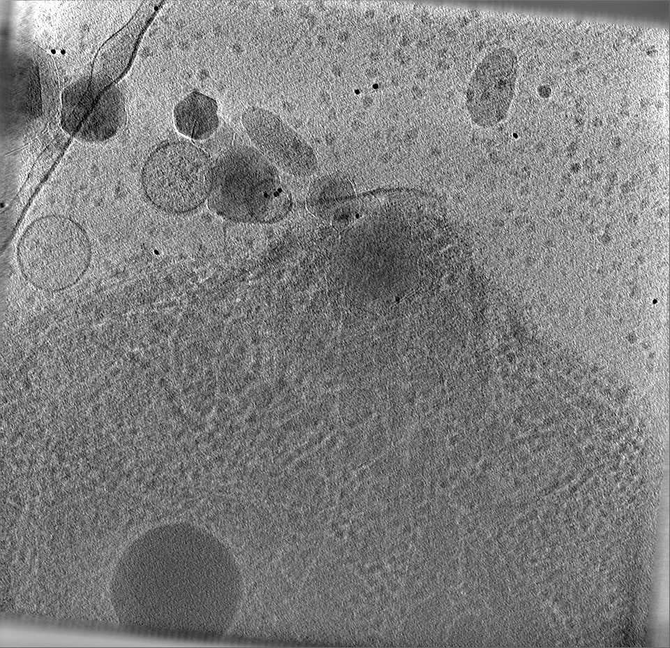
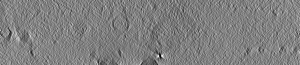
Tachyzoites were released from heavily infected monolayers of HFFs by mechanical disruption of the monolayers using disposable scrapers and passage through a 25-gauge syringe into HPEB
Sectioning
Other: NO SECTIONING
Fiducial marker
Manufacturer: EMS / Diameter: 10 nm
-
Electron microscopy
Microscope
TFS KRIOS
Specialist optics
Phase plate: VOLTA PHASE PLATE / Energy filter - Name: GIF Bioquantum
Image recording
Film or detector model: GATAN K2 SUMMIT (4k x 4k) / Average electron dose: 1.0 e/Å2
Electron beam
Acceleration voltage: 300 kV / Electron source: FIELD EMISSION GUN
In the structure databanks used in Yorodumi, some data are registered as the other names, "COVID-19 virus" and "2019-nCoV". Here are the details of the virus and the list of structure data.
Jan 31, 2019. EMDB accession codes are about to change! (news from PDBe EMDB page)
EMDB accession codes are about to change! (news from PDBe EMDB page)
The allocation of 4 digits for EMDB accession codes will soon come to an end. Whilst these codes will remain in use, new EMDB accession codes will include an additional digit and will expand incrementally as the available range of codes is exhausted. The current 4-digit format prefixed with “EMD-” (i.e. EMD-XXXX) will advance to a 5-digit format (i.e. EMD-XXXXX), and so on. It is currently estimated that the 4-digit codes will be depleted around Spring 2019, at which point the 5-digit format will come into force.
The EM Navigator/Yorodumi systems omit the EMD- prefix.
Related info.:Q: What is EMD? / ID/Accession-code notation in Yorodumi/EM Navigator
Yorodumi is a browser for structure data from EMDB, PDB, SASBDB, etc.
This page is also the successor to EM Navigator detail page, and also detail information page/front-end page for Omokage search.
The word "yorodu" (or yorozu) is an old Japanese word meaning "ten thousand". "mi" (miru) is to see.
Related info.:EMDB / PDB / SASBDB / Comparison of 3 databanks / Yorodumi Search / Aug 31, 2016. New EM Navigator & Yorodumi / Yorodumi Papers / Jmol/JSmol / Function and homology information / Changes in new EM Navigator and Yorodumi
 Movie
Movie Controller


 Open data
 Basic information
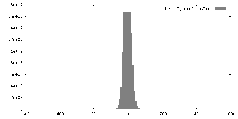
 Map data
 Sample
 Keywords
 Authors
 United States, 2 items
  Citation
 Structure visualization
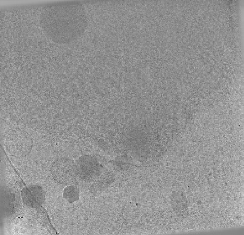
 Downloads & links
 EMDB map data format
 emd_28140.png
 http://ftp.pdbj.org/pub/emdb/structures/EMD-28140
 Z (Sec.)
 Y (Row.)
 X (Col.)


 Sample components
 Processing
 Electron microscopy
 FIELD EMISSION GUN
