Movie
Movie Controller

[English] 日本語
 Yorodumi
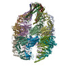
- PDB-8i6j: The focused refinement of CCT3-PhLP2A from TRiC-PhLP2A complex in... -
+ Open data

- Basic information
| Entry | Database: PDB / ID: 8i6j | ||||||
|---|---|---|---|---|---|---|---|
| Title | The focused refinement of CCT3-PhLP2A from TRiC-PhLP2A complex in the open state | ||||||
 Components Components |
| ||||||
 Keywords Keywords | CHAPERONE / chaperonin complex / cochaperone | ||||||
| Function / homology |  Function and homology information Function and homology informationnegative regulation of protein folding / perinucleolar compartment / positive regulation of protein localization to Cajal body / zona pellucida receptor complex / positive regulation of telomerase RNA localization to Cajal body / chaperonin-containing T-complex / BBSome-mediated cargo-targeting to cilium / Formation of tubulin folding intermediates by CCT/TriC / binding of sperm to zona pellucida / vascular endothelial growth factor receptor 2 binding ...negative regulation of protein folding / perinucleolar compartment / positive regulation of protein localization to Cajal body / zona pellucida receptor complex / positive regulation of telomerase RNA localization to Cajal body / chaperonin-containing T-complex / BBSome-mediated cargo-targeting to cilium / Formation of tubulin folding intermediates by CCT/TriC / binding of sperm to zona pellucida / vascular endothelial growth factor receptor 2 binding / Folding of actin by CCT/TriC / Prefoldin mediated transfer of substrate to CCT/TriC / regulation of peptidyl-tyrosine phosphorylation / Association of TriC/CCT with target proteins during biosynthesis / negative regulation of ubiquitin-dependent protein catabolic process / Hydrolases; Acting on acid anhydrides; In phosphorus-containing anhydrides / positive regulation of telomere maintenance via telomerase / positive regulation of endothelial cell proliferation / protein folding chaperone / ATP-dependent protein folding chaperone / positive regulation of angiogenesis / : / Cooperation of PDCL (PhLP1) and TRiC/CCT in G-protein beta folding / protein folding / actin cytoskeleton organization / cell body / angiogenesis / cytoskeleton / microtubule / protein stabilization / apoptotic process / positive regulation of gene expression / perinuclear region of cytoplasm / endoplasmic reticulum / ATP hydrolysis activity / protein-containing complex / RNA binding / extracellular exosome / nucleoplasm / ATP binding / cytoplasm / cytosol Similarity search - Function | ||||||
| Biological species |  Homo sapiens (human) Homo sapiens (human) | ||||||
| Method | ELECTRON MICROSCOPY / single particle reconstruction / cryo EM / Resolution: 3.82 Å | ||||||
 Authors Authors | Roh, S.H. / Park, J. / Kim, H. / Lim, S. | ||||||
| Funding support |  Korea, Republic Of, 1items Korea, Republic Of, 1items
| ||||||
 Citation Citation | Journal: Nat Commun / Year: 2024 Title: A structural vista of phosducin-like PhLP2A-chaperonin TRiC cooperation during the ATP-driven folding cycle. Authors: Junsun Park / Hyunmin Kim / Daniel Gestaut / Seyeon Lim / Kwadwo A Opoku-Nsiah / Alexander Leitner / Judith Frydman / Soung-Hun Roh /   Abstract: Proper cellular proteostasis, essential for viability, requires a network of chaperones and cochaperones. ATP-dependent chaperonin TRiC/CCT partners with cochaperones prefoldin (PFD) and phosducin- ...Proper cellular proteostasis, essential for viability, requires a network of chaperones and cochaperones. ATP-dependent chaperonin TRiC/CCT partners with cochaperones prefoldin (PFD) and phosducin-like proteins (PhLPs) to facilitate folding of essential eukaryotic proteins. Using cryoEM and biochemical analyses, we determine the ATP-driven cycle of TRiC-PFD-PhLP2A interaction. PhLP2A binds to open apo-TRiC through polyvalent domain-specific contacts with its chamber's equatorial and apical regions. PhLP2A N-terminal H3-domain binding to subunits CCT3/4 apical domains displace PFD from TRiC. ATP-induced TRiC closure rearranges the contacts of PhLP2A domains within the closed chamber. In the presence of substrate, actin and PhLP2A segregate into opposing chambers, each binding to positively charged inner surface residues from CCT1/3/6/8. Notably, actin induces a conformational change in PhLP2A, causing its N-terminal helices to extend across the inter-ring interface to directly contact a hydrophobic groove in actin. Our findings reveal an ATP-driven PhLP2A structural rearrangement cycle within the TRiC chamber to facilitate folding. | ||||||
| History |
|
- Structure visualization
| Structure viewer | Molecule: MolmilJmol/JSmol |
|---|
- Downloads & links
-Download
| PDBx/mmCIF format | 8i6j.cif.gz | 132.1 KB | Display | PDBx/mmCIF format |
|---|---|---|---|---|
| PDB format | pdb8i6j.ent.gz | 99.7 KB | Display | PDB format |
| PDBx/mmJSON format | 8i6j.json.gz | Tree view | PDBx/mmJSON format | |
| Others |  Other downloads Other downloads |
-Validation report
| Arichive directory | https://data.pdbj.org/pub/pdb/validation_reports/i6/8i6jftp://data.pdbj.org/pub/pdb/validation_reports/i6/8i6j | HTTPS FTP |
|---|
-Related structure data
| Related structure data |  35199MC  8i1uC  8i9qC  8i9uC  8ib8C M: map data used to model this data C: citing same article ( |
|---|---|
| Similar structure data |


-Links
 PDBj
PDBj


- Assembly
| Deposited unit | 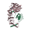
|
|---|---|
| 1 |
|
-Components
| #1: Protein | Mass: 27650.383 Da / Num. of mol.: 1 Source method: isolated from a genetically manipulated source Source: (gene. exp.) Homo sapiens (human) / Gene: PDCL3, PhLP2A / Production host:  |
|---|---|
| #2: Protein | Mass: 60613.855 Da / Num. of mol.: 1 Source method: isolated from a genetically manipulated source Source: (gene. exp.) Homo sapiens (human) / Gene: CCT3, CCTG, TRIC5 / Cell line (production host): High Five / Production host:  Trichoplusia ni (cabbage looper) / References: UniProt: P49368 Trichoplusia ni (cabbage looper) / References: UniProt: P49368 |
| #3: Chemical | ChemComp-ADP /   Mass: 427.201 Da / Num. of mol.: 1 / Source method: obtained synthetically / Formula: C10H15N5O10P2 / Feature type: SUBJECT OF INVESTIGATION / Comment: ADP, energy-carrying molecule*YM Mass: 427.201 Da / Num. of mol.: 1 / Source method: obtained synthetically / Formula: C10H15N5O10P2 / Feature type: SUBJECT OF INVESTIGATION / Comment: ADP, energy-carrying molecule*YM |
| Has ligand of interest | Y |
-Experimental details
-Experiment
| Experiment | Method: ELECTRON MICROSCOPY |
|---|---|
| EM experiment | Aggregation state: 2D ARRAY / 3D reconstruction method: single particle reconstruction |
- Sample preparation
| Component |
| ||||||||||||||||||||||||
|---|---|---|---|---|---|---|---|---|---|---|---|---|---|---|---|---|---|---|---|---|---|---|---|---|---|
| Molecular weight |
| ||||||||||||||||||||||||
| Source (natural) |
| ||||||||||||||||||||||||
| Source (recombinant) |
| ||||||||||||||||||||||||
| Buffer solution | pH: 7.4 | ||||||||||||||||||||||||
| Buffer component |
| ||||||||||||||||||||||||
| Specimen | Embedding applied: NO / Shadowing applied: NO / Staining applied: NO / Vitrification applied: YES | ||||||||||||||||||||||||
| Specimen support | Grid type: Quantifoil R1.2/1.3 | ||||||||||||||||||||||||
| Vitrification | Instrument: FEI VITROBOT MARK IV / Cryogen name: ETHANE / Humidity: 100 % |
- Electron microscopy imaging
| Experimental equipment |  Model: Titan Krios / Image courtesy: FEI Company |
|---|---|
| Microscopy | Model: FEI TITAN KRIOS |
| Electron gun | Electron source:  FIELD EMISSION GUN / Accelerating voltage: 300 kV / Illumination mode: FLOOD BEAM FIELD EMISSION GUN / Accelerating voltage: 300 kV / Illumination mode: FLOOD BEAM |
| Electron lens | Mode: BRIGHT FIELD / Nominal defocus max: 2000 nm / Nominal defocus min: 1000 nm / Cs: 2.7 mm |
| Specimen holder | Cryogen: NITROGEN / Specimen holder model: FEI TITAN KRIOS AUTOGRID HOLDER |
| Image recording | Electron dose: 50 e/Å2 / Detector mode: COUNTING / Film or detector model: FEI FALCON IV (4k x 4k) / Num. of real images: 15075 |
| Image scans | Width: 4096 / Height: 4096 |
- Processing
| Software | Name: PHENIX / Version: 1.19.2_4158: / Classification: refinement | ||||||||||||||||||||||||||||||||||||||||||||||||||
|---|---|---|---|---|---|---|---|---|---|---|---|---|---|---|---|---|---|---|---|---|---|---|---|---|---|---|---|---|---|---|---|---|---|---|---|---|---|---|---|---|---|---|---|---|---|---|---|---|---|---|---|
| EM software |
| ||||||||||||||||||||||||||||||||||||||||||||||||||
| CTF correction | Details: CTF correction was performed for every micrographs / Type: NONE | ||||||||||||||||||||||||||||||||||||||||||||||||||
| Particle selection | Num. of particles selected: 2691733 Details: The initial particle selection after 2D class classification | ||||||||||||||||||||||||||||||||||||||||||||||||||
| 3D reconstruction | Resolution: 3.82 Å / Resolution method: FSC 0.143 CUT-OFF / Num. of particles: 359533 / Details: Focused refinement map for CCT3 and PhLP2A complex / Symmetry type: POINT | ||||||||||||||||||||||||||||||||||||||||||||||||||
| Atomic model building | Protocol: FLEXIBLE FIT / Space: REAL | ||||||||||||||||||||||||||||||||||||||||||||||||||
| Atomic model building | PDB-ID: 6NR8 Accession code: 6NR8 / Source name: PDB / Type: experimental model | ||||||||||||||||||||||||||||||||||||||||||||||||||
| Refine LS restraints |
|