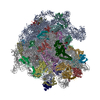
 ムービー
ムービー コントローラー
 構造ビューア
 EMN検索について


-検索条件
-検索結果
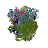
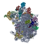
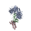
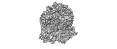
検索 (著者・登録者: sone & k)の結果55件中、1から50件目までを表示しています
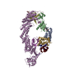
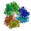
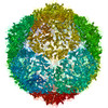
PDB-8rx0: 
(NEDD8)-CRL2VHL-MZ1-Brd4BD2-Ub(G76S, K48C)-UBE2R1(C93K, S138C, C191S, C223S)-Ub
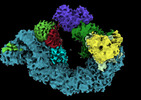
EMDB-19567:
Closed crosslinked structure of (NEDD8)-CRL2VHL-MZ1-Brd4BD2-Ub(G76S, K48C)-UBE2R1(C93K, S138C, C191S, C223S)-Ub

EMDB-19569:
Open non-crosslinked structure Brd4BD2-MZ1-(NEDD8)-CRL2VHL

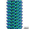
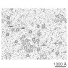
EMDB-18004:
Cryo electron tomogram of Caulobacter crescentus - Delta-bla

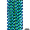
EMDB-18005:
Cryo electron tomogram of Caulobacter crescentus - Delta-bla;tipT::Tn

EMDB-18006:
Cryo electron tomogram of Caulobacter crescentus - Delta-bla;pSRKacrAB::nodT

EMDB-18007:
Cryo electron tomogram of Caulobacter crescentus - Delta-bla;pSRKacrAB::nodT example 2

EMDB-18008:
Cryo electron tomogram of Caulobacter crescentus - Delta-bla;tipR::Tn
EMDB-29298:
The structure of a hibernating ribosome in the Lyme disease pathogen
EMDB-29304:
The structure of a 50S ribosomal subunit in the Lyme disease pathogen Borreliella burgdorferi
PDB-8fmw:
The structure of a hibernating ribosome in the Lyme disease pathogen
PDB-8fn2:
The structure of a 50S ribosomal subunit in the Lyme disease pathogen Borreliella burgdorferi

EMDB-17172:
Ternary structure of intramolecular bivalent glue degrader IBG1 bound to BRD4 and DCAF16:DDB1deltaBPB
PDB-8ov6:
Ternary structure of intramolecular bivalent glue degrader IBG1 bound to BRD4 and DCAF16:DDB1deltaBPB
EMDB-29397:
Structure of Mycobacterium smegmatis Rsh bound to a 70S translation initiation complex
PDB-8fr8:
Structure of Mycobacterium smegmatis Rsh bound to a 70S translation initiation complex
EMDB-16226:
Giardia Ribosome in PRE-T Classical State (C)
EMDB-16228:
Giardia Ribosome in PRE-T Hybrid State (D1)
EMDB-16235:
Giardia Ribosome in PRE-T Hybrid State (D2)

PDB-8bsj:
Giardia Ribosome in PRE-T Classical State (C)

PDB-8btd:
Giardia Ribosome in PRE-T Hybrid State (D1)

PDB-8btr:
Giardia Ribosome in PRE-T Hybrid State (D2)
EMDB-16211:
Giardia ribosome in POST-T state (A1)
EMDB-16222:
Giardia ribosome in POST-T state, no E-site tRNA (A6)
EMDB-16225:
Giardia ribosome chimeric hybrid-like GDP+Pi bound state (B1)

PDB-8br8:
Giardia ribosome in POST-T state (A1)

PDB-8brm:
Giardia ribosome in POST-T state, no E-site tRNA (A6)

PDB-8bsi:
Giardia ribosome chimeric hybrid-like GDP+Pi bound state (B1)
EMDB-30440:
Cryo-EM analysis of the nonribosomal peptide synthetase, FmoA3
EMDB-23211:
Cryo-EM structure of human ACE2 receptor bound to protein encoded by vaccine candidate BNT162b1
EMDB-23215:
Cryo-EM structure of protein encoded by vaccine candidate BNT162b2

PDB-7l7f:
Cryo-EM structure of human ACE2 receptor bound to protein encoded by vaccine candidate BNT162b1

PDB-7l7k:
Cryo-EM structure of protein encoded by vaccine candidate BNT162b2
EMDB-8166:
Structure of the S. cerevisiae alpha-mannosidase 1
EMDB-8167:
Structure of S. cerevesiae mApe1 dodecamer

PDB-5jm0:
Structure of the S. cerevisiae alpha-mannosidase 1
PDB-5jm9:
Structure of S. cerevesiae mApe1 dodecamer
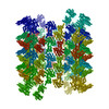
EMDB-2896:
Cryo-EM structure of helical ANTH and ENTH tubules on PI(4,5)P2-containing membranes
EMDB-2897:
Cryo-EM structure of helical ANTH and ENTH tubules on PI(4,5)P2-containing membranes
PDB-5ahv:
Cryo-EM structure of helical ANTH and ENTH tubules on PI(4,5)P2-containing membranes
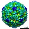
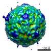
PDB-4bip:
Homology model of coxsackievirus A7 (CAV7) full capsid proteins.

PDB-4biq:
Homology model of coxsackievirus A7 (CAV7) empty capsid proteins.
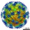
EMDB-5512:
Icosahedral reconstruction of filled coxsackievirus A9 capsid-integrin alpha v beta 6 complex
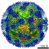
EMDB-5514:
Icosahedral reconstruction of empty coxsackievirus A9 capsid-integrin alpha v beta 6 complex
EMDB-5515:
Icosahedral reconstruction of filled coxsackievirus A9 capsid
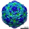
EMDB-5516:
Icosahedral reconstruction of empty coxsackievirus A9 capsid
EMDB-5517:
Asymmetric reconstruction of filled-integrin alpha v beta 6 complex
EMDB-5519:
Electron cryo-tomography of coxsackievirus A9-integrin alpha v beta 6 complex
PDB-3j2j:
Empty coxsackievirus A9 capsid
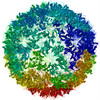
EMDB-2027:
Coxsackievirus A7 (CAV7) empty capsid reconstruction at 6.09 angstrom resolution.
ページ:
 wwPDBはEMDBデータモデルのバージョン3へ移行します
