 Movie
Movie Controller


+ Open data

- Basic information
| Entry | Database: PDB / ID: 5jm0 | ||||||
|---|---|---|---|---|---|---|---|
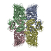
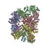
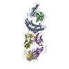
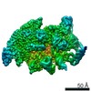
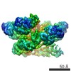
| Title | Structure of the S. cerevisiae alpha-mannosidase 1 | ||||||
 Components Components | Alpha-mannosidase,Alpha-mannosidase,Alpha-mannosidase | ||||||
 Keywords Keywords | HYDROLASE / tetramer / cvt cargo / mannosidase / selective autophagy | ||||||
| Function / homology |  Function and homology information Function and homology informationmannose catabolic process / Lysosomal oligosaccharide catabolism / Cvt complex / alpha-mannosidase / alpha-mannosidase activity / fungal-type vacuole membrane / oligosaccharide catabolic process / cellular response to nitrogen starvation / cellular response to glucose starvation / carbohydrate binding / metal ion binding Similarity search - Function | ||||||
| Biological species |  | ||||||
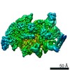
| Method | ELECTRON MICROSCOPY / single particle reconstruction / cryo EM / Resolution: 6.3 Å | ||||||
 Authors Authors | Schneider, S. / Kosinski, J. / Jakobi, A.J. / Hagen, W.J.H. / Sachse, C. | ||||||
 Citation Citation | Journal: EMBO Rep / Year: 2016 Title: Higher-order assemblies of oligomeric cargo receptor complexes form the membrane scaffold of the Cvt vesicle. Authors: Chiara Bertipaglia / Sarah Schneider / Arjen J Jakobi / Abul K Tarafder / Yury S Bykov / Andrea Picco / Wanda Kukulski / Jan Kosinski / Wim Jh Hagen / Arvind C Ravichandran / Matthias ...Authors: Chiara Bertipaglia / Sarah Schneider / Arjen J Jakobi / Abul K Tarafder / Yury S Bykov / Andrea Picco / Wanda Kukulski / Jan Kosinski / Wim Jh Hagen / Arvind C Ravichandran / Matthias Wilmanns / Marko Kaksonen / John Ag Briggs / Carsten Sachse /  Abstract: Selective autophagy is the mechanism by which large cargos are specifically sequestered for degradation. The structural details of cargo and receptor assembly giving rise to autophagic vesicles ...Selective autophagy is the mechanism by which large cargos are specifically sequestered for degradation. The structural details of cargo and receptor assembly giving rise to autophagic vesicles remain to be elucidated. We utilize the yeast cytoplasm-to-vacuole targeting (Cvt) pathway, a prototype of selective autophagy, together with a multi-scale analysis approach to study the molecular structure of Cvt vesicles. We report the oligomeric nature of the major Cvt cargo Ape1 with a combined 2.8 Å X-ray and negative stain EM structure, as well as the secondary cargo Ams1 with a 6.3 Å cryo-EM structure. We show that the major dodecameric cargo prApe1 exhibits a tendency to form higher-order chain structures that are broken upon interaction with the receptor Atg19 in vitro The stoichiometry of these cargo-receptor complexes is key to maintaining the size of the Cvt aggregate in vivo Using correlative light and electron microscopy, we further visualize key stages of Cvt vesicle biogenesis. Our findings suggest that Atg19 interaction limits Ape1 aggregate size while serving as a vehicle for vacuolar delivery of tetrameric Ams1. | ||||||
| History |
|
- Structure visualization
| Movie |
Movie viewer |
|---|---|
| Structure viewer | Molecule: MolmilJmol/JSmol |
- Downloads & links
-Download
| PDBx/mmCIF format | 5jm0.cif.gz | 187.5 KB | Display | PDBx/mmCIF format |
|---|---|---|---|---|
| PDB format | pdb5jm0.ent.gz | 144.1 KB | Display | PDB format |
| PDBx/mmJSON format | 5jm0.json.gz | Tree view | PDBx/mmJSON format | |
| Others |  Other downloads Other downloads |
-Validation report
| Arichive directory | https://data.pdbj.org/pub/pdb/validation_reports/jm/5jm0ftp://data.pdbj.org/pub/pdb/validation_reports/jm/5jm0 | HTTPS FTP |
|---|
-Related structure data
| Related structure data | 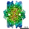 8166MC 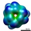 8167C 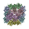 5jm6C 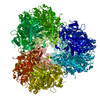 5jm9C M: map data used to model this data C: citing same article ( |
|---|---|
| Similar structure data |

-Links
 PDBj
PDBj


- Assembly
| Deposited unit | 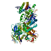
|
|---|---|
| 1 | 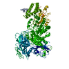
|
| Symmetry | Point symmetry: (Schoenflies symbol: D2 (2x2 fold dihedral)) |
-Components
| #1: Protein | Mass: 125764.156 Da / Num. of mol.: 1 Source method: isolated from a genetically manipulated source Details: The coordinate model contains a poly-alanine stretch that corresponds to residues 17-27 in the template sequence. Source: (gene. exp.)  Komagataella pastoris (fungus) / Strain (production host): SMD1168 / References: UniProt: P22855, alpha-mannosidase Komagataella pastoris (fungus) / Strain (production host): SMD1168 / References: UniProt: P22855, alpha-mannosidase |
|---|
-Experimental details
-Experiment
| Experiment | Method: ELECTRON MICROSCOPY |
|---|---|
| EM experiment | Aggregation state: PARTICLE / 3D reconstruction method: single particle reconstruction |
- Sample preparation
| Component | Name: Alpha-mannosidase 1 / Type: COMPLEX / Details: His-tagged / Entity ID: all / Source: RECOMBINANT | ||||||||||||||||||||
|---|---|---|---|---|---|---|---|---|---|---|---|---|---|---|---|---|---|---|---|---|---|
| Molecular weight | Value: 0.5 MDa / Experimental value: NO | ||||||||||||||||||||
| Source (natural) | Organism: | ||||||||||||||||||||
| Source (recombinant) | Organism: Komagataella pastoris (fungus) / Strain: SMD1168 / Plasmid: pGAPz | ||||||||||||||||||||
| Buffer solution | pH: 7.5 | ||||||||||||||||||||
| Buffer component |
| ||||||||||||||||||||
| Specimen | Conc.: 0.4 mg/ml / Embedding applied: NO / Shadowing applied: NO / Staining applied: NO / Vitrification applied: YES / Details: single particles alongside chains of tetramers | ||||||||||||||||||||
| Specimen support | Grid material: COPPER / Grid mesh size: 300 divisions/in. / Grid type: Quantifoil R2/2 | ||||||||||||||||||||
| Vitrification | Instrument: FEI VITROBOT MARK III / Cryogen name: ETHANE / Humidity: 95 % / Chamber temperature: 293 K Details: 2.5 ul of sample was applied, offset -3, 9s blotting time |
- Electron microscopy imaging
| Experimental equipment |  Model: Titan Krios / Image courtesy: FEI Company |
|---|---|
| Microscopy | Model: FEI TITAN KRIOS |
| Electron gun | Electron source:  FIELD EMISSION GUN / Accelerating voltage: 300 kV / Illumination mode: FLOOD BEAM FIELD EMISSION GUN / Accelerating voltage: 300 kV / Illumination mode: FLOOD BEAM |
| Electron lens | Mode: BRIGHT FIELD / Nominal magnification: 75000 X / Nominal defocus max: 5000 nm / Nominal defocus min: 1000 nm / Cs: 2.7 mm / C2 aperture diameter: 50 µm / Alignment procedure: COMA FREE |
| Specimen holder | Cryogen: NITROGEN / Specimen holder model: FEI TITAN KRIOS AUTOGRID HOLDER |
| Image recording | Average exposure time: 1.6 sec. / Electron dose: 58 e/Å2 / Detector mode: INTEGRATING / Film or detector model: FEI FALCON II (4k x 4k) / Num. of grids imaged: 1 / Num. of real images: 1735 Details: The first six frames comprised each 7 e/A2 and the final frame consequently received 16 e/A2 dose. Relion's particle polishing procedure was used. |
| Image scans | Movie frames/image: 7 / Used frames/image: 1-7 |
- Processing
| EM software |
| |||||||||||||||||||||||||||||||||||||||||||||||||||||||
|---|---|---|---|---|---|---|---|---|---|---|---|---|---|---|---|---|---|---|---|---|---|---|---|---|---|---|---|---|---|---|---|---|---|---|---|---|---|---|---|---|---|---|---|---|---|---|---|---|---|---|---|---|---|---|---|---|
| CTF correction | Type: PHASE FLIPPING AND AMPLITUDE CORRECTION | |||||||||||||||||||||||||||||||||||||||||||||||||||||||
| Particle selection | Num. of particles selected: 85274 | |||||||||||||||||||||||||||||||||||||||||||||||||||||||
| Symmetry | Point symmetry: D2 (2x2 fold dihedral) | |||||||||||||||||||||||||||||||||||||||||||||||||||||||
| 3D reconstruction | Resolution: 6.3 Å / Resolution method: FSC 0.143 CUT-OFF / Num. of particles: 33588 / Algorithm: FOURIER SPACE Details: 1. From 85274 initially auto-picked particles, 16678 were eliminated after 2D classification. 2. From 68596, two classes making up 33588 particles were included in the final reconstruction. Num. of class averages: 2 / Symmetry type: POINT | |||||||||||||||||||||||||||||||||||||||||||||||||||||||
| Atomic model building | B value: 80 / Protocol: FLEXIBLE FIT / Space: REAL / Target criteria: Cross-correlation coefficient Details: 1. Fitting of a homology model based on PDB 2wyh combined with a homology model based on PDB 2xbz and 3cmg for the C- (residues 287-1083) and N-terminal part (residues 45-203) respectively 2. ...Details: 1. Fitting of a homology model based on PDB 2wyh combined with a homology model based on PDB 2xbz and 3cmg for the C- (residues 287-1083) and N-terminal part (residues 45-203) respectively 2. ab initio modelling for a three-helix bundle of the N-terminal part (residues 209-286) 3. creating an ideal poly-alanine helix for residues 17-27 |
