 Movie
Movie Controller


+ Open data

- Basic information
| Entry | Database: PDB / ID: 6lz1 | ||||||||||||
|---|---|---|---|---|---|---|---|---|---|---|---|---|---|
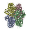
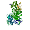
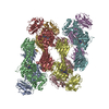
| Title | Structure of S.pombe alpha-mannosidase Ams1 | ||||||||||||
 Components Components | Ams1 | ||||||||||||
 Keywords Keywords | HYDROLASE / glycoside hydrolase | ||||||||||||
| Function / homology |  Function and homology information Function and homology informationmannosyl-oligosaccharide 1,6-alpha-mannosidase activity / mannosyl-oligosaccharide 1,3-alpha-mannosidase activity / Lysosomal oligosaccharide catabolism / fungal-type vacuole lumen / alpha-mannosidase / alpha-mannosidase activity / mannose metabolic process / fungal-type vacuole membrane / oligosaccharide catabolic process / carbohydrate binding ...mannosyl-oligosaccharide 1,6-alpha-mannosidase activity / mannosyl-oligosaccharide 1,3-alpha-mannosidase activity / Lysosomal oligosaccharide catabolism / fungal-type vacuole lumen / alpha-mannosidase / alpha-mannosidase activity / mannose metabolic process / fungal-type vacuole membrane / oligosaccharide catabolic process / carbohydrate binding / metal ion binding / cytosol Similarity search - Function | ||||||||||||
| Biological species |  | ||||||||||||
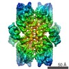
| Method | ELECTRON MICROSCOPY / single particle reconstruction / cryo EM / Resolution: 3.2 Å | ||||||||||||
 Authors Authors | Zhang, J. / Ye, K. | ||||||||||||
| Funding support |  China, 3items China, 3items
| ||||||||||||
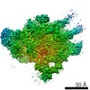
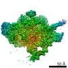
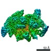
 Citation Citation | Journal: FEBS Open Bio / Year: 2020 Title: Cryo-EM structure of fission yeast tetrameric α-mannosidase Ams1. Authors: Jianxiu Zhang / Ying-Ying Wang / Li-Lin Du / Keqiong Ye / Abstract: Fungal α-mannosidase Ams1 and its mammalian homolog MAN2C1 hydrolyze terminal α-linked mannoses in free oligosaccharides released from misfolded glycoproteins or lipid-linked oligosaccharide donors. ...Fungal α-mannosidase Ams1 and its mammalian homolog MAN2C1 hydrolyze terminal α-linked mannoses in free oligosaccharides released from misfolded glycoproteins or lipid-linked oligosaccharide donors. Ams1 is transported by selective autophagy into vacuoles. Here, we determine the tetrameric structure of Ams1 from the fission yeast Schizosaccharomyces pombe at 3.2 Å resolution by cryo-electron microscopy. Distinct from a low resolution structure of S. cerevisiae Ams1, S. pombe Ams1 has a prominent N-terminal tail that mediates tetramerization and an extra β-sheet domain. Ams1 shares a conserved active site with other enzymes in glycoside hydrolase family 38, to which Ams1 belongs, but contains extra N-terminal domains involved in tetramerization. The atomic structure of Ams1 reported here will aid understanding of its enzymatic activity and transport mechanism. | ||||||||||||
| History |
|
- Structure visualization
| Movie |
Movie viewer |
|---|---|
| Structure viewer | Molecule: MolmilJmol/JSmol |
- Downloads & links
-Download
| PDBx/mmCIF format | 6lz1.cif.gz | 752.2 KB | Display | PDBx/mmCIF format |
|---|---|---|---|---|
| PDB format | pdb6lz1.ent.gz | 614.2 KB | Display | PDB format |
| PDBx/mmJSON format | 6lz1.json.gz | Tree view | PDBx/mmJSON format | |
| Others |  Other downloads Other downloads |
-Validation report
| Arichive directory | https://data.pdbj.org/pub/pdb/validation_reports/lz/6lz1ftp://data.pdbj.org/pub/pdb/validation_reports/lz/6lz1 | HTTPS FTP |
|---|
-Related structure data
| Related structure data |  30021MC M: map data used to model this data C: citing same article ( |
|---|---|
| Similar structure data |

-Links
 PDBj
PDBj


- Assembly
| Deposited unit | 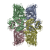
|
|---|---|
| 1 |
|
-Components
| #1: Protein | Mass: 129201.672 Da / Num. of mol.: 4 / Fragment: Znic ion Source method: isolated from a genetically manipulated source Source: (gene. exp.) Gene: ams1, mns2, SPAC513.05 / Production host: #2: Chemical | ChemComp-ZN /   Mass: 65.409 Da / Num. of mol.: 4 / Source method: obtained synthetically / Formula: Zn / Feature type: SUBJECT OF INVESTIGATION Mass: 65.409 Da / Num. of mol.: 4 / Source method: obtained synthetically / Formula: Zn / Feature type: SUBJECT OF INVESTIGATIONHas ligand of interest | Y | |
|---|
-Experimental details
-Experiment
| Experiment | Method: ELECTRON MICROSCOPY |
|---|---|
| EM experiment | Aggregation state: PARTICLE / 3D reconstruction method: single particle reconstruction |
- Sample preparation
| Component | Name: Ams1 tetramer / Type: COMPLEX / Entity ID: #1 / Source: RECOMBINANT | |||||||||||||||||||||||||
|---|---|---|---|---|---|---|---|---|---|---|---|---|---|---|---|---|---|---|---|---|---|---|---|---|---|---|
| Molecular weight | Value: 0.48 MDa / Experimental value: NO | |||||||||||||||||||||||||
| Source (natural) | Organism: | |||||||||||||||||||||||||
| Source (recombinant) | Organism: | |||||||||||||||||||||||||
| Buffer solution | pH: 7.5 | |||||||||||||||||||||||||
| Buffer component |
| |||||||||||||||||||||||||
| Specimen | Conc.: 0.5 mg/ml / Embedding applied: NO / Shadowing applied: NO / Staining applied: NO / Vitrification applied: YES | |||||||||||||||||||||||||
| Specimen support | Grid material: COPPER / Grid mesh size: 300 divisions/in. / Grid type: Quantifoil R1.2/1.3 | |||||||||||||||||||||||||
| Vitrification | Instrument: FEI VITROBOT MARK IV / Cryogen name: ETHANE / Humidity: 100 % / Chamber temperature: 277 K / Details: blot for 5 seconds before plunging |
- Electron microscopy imaging
| Experimental equipment |  Model: Titan Krios / Image courtesy: FEI Company |
|---|---|
| Microscopy | Model: FEI TITAN KRIOS |
| Electron gun | Electron source:  FIELD EMISSION GUN / Accelerating voltage: 300 kV / Illumination mode: FLOOD BEAM FIELD EMISSION GUN / Accelerating voltage: 300 kV / Illumination mode: FLOOD BEAM |
| Electron lens | Mode: BRIGHT FIELD / Nominal defocus max: 1000 nm / Nominal defocus min: 400 nm / Cs: 2.7 mm |
| Specimen holder | Cryogen: NITROGEN / Specimen holder model: FEI TITAN KRIOS AUTOGRID HOLDER |
| Image recording | Average exposure time: 8.4 sec. / Electron dose: 60 e/Å2 / Detector mode: SUPER-RESOLUTION / Film or detector model: GATAN K2 SUMMIT (4k x 4k) / Num. of real images: 597 |
| EM imaging optics | Energyfilter name: GIF Tridiem 4K / Energyfilter slit width: 20 eV / Phase plate: VOLTA PHASE PLATE |
| Image scans | Movie frames/image: 32 |
- Processing
| Software |
| ||||||||||||||||||||||||||||||||||||||||
|---|---|---|---|---|---|---|---|---|---|---|---|---|---|---|---|---|---|---|---|---|---|---|---|---|---|---|---|---|---|---|---|---|---|---|---|---|---|---|---|---|---|
| EM software |
| ||||||||||||||||||||||||||||||||||||||||
| CTF correction | Type: PHASE FLIPPING AND AMPLITUDE CORRECTION | ||||||||||||||||||||||||||||||||||||||||
| Symmetry | Point symmetry: D2 (2x2 fold dihedral) | ||||||||||||||||||||||||||||||||||||||||
| 3D reconstruction | Resolution: 3.2 Å / Resolution method: FSC 0.143 CUT-OFF / Num. of particles: 75460 / Symmetry type: POINT | ||||||||||||||||||||||||||||||||||||||||
| Atomic model building | Protocol: AB INITIO MODEL / Space: REAL / Target criteria: Correlation coefficient | ||||||||||||||||||||||||||||||||||||||||
| Atomic model building | PDB-ID: 6LZ1 Accession code: 6LZ1 / Source name: PDB / Type: experimental model | ||||||||||||||||||||||||||||||||||||||||
| Refinement | Cross valid method: NONE Stereochemistry target values: GeoStd + Monomer Library + CDL v1.2 | ||||||||||||||||||||||||||||||||||||||||
| Displacement parameters | Biso mean: 31.48 Å2 | ||||||||||||||||||||||||||||||||||||||||
| Refine LS restraints |
|
