 Movie
Movie Controller
 Structure viewers
 About EMN search


-Search query
-Search result
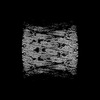
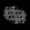
Showing 1 - 50 of 114 items for (author: roux & a)

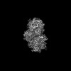
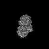
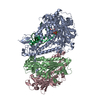
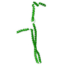
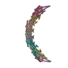
EMDB-56110: 
Flat clathrin lattice on endosomes
Method: subtomogram averaging / : Gul M, Hakala M, Moparthi SB, Ganeva I, Bernat-Silvestre C, Marcuello C, Espadas J, Colom A, Kukulski W, Vassilopoulos S, Kaksonen M, Roux A, Kudryashev M

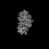
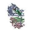
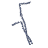
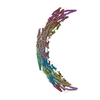
EMDB-56112:
Cryo-electron tomogram of endosomes in HeLa cells
Method: electron tomography / : Hakala M, Moparthi SB, Ganeva I, Gul M, Bernat-Silvestre C, Marcuello C, Espadas J, Colom A, Kudryashev M, Kukulski W, Vassilopoulos S, Kaksonen M, Roux A
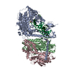
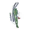
EMDB-51326:
Structure of the G848S mutant of human mitochondrial DNA polymerase gamma in complex with PZL-A
Method: single particle / : Valenzuela S, Falkenberg M
EMDB-51327:
Structure of the G848S mutant of human mitochondrial DNA polymerase gamma
Method: single particle / : Valenzuela S, Falkenberg M
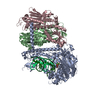
EMDB-51328:
Structure of the A467T mutant of human mitochondrial DNA polymerase gamma in complex with PZL-A
Method: single particle / : Valenzuela S, Falkenberg M
EMDB-51329:
Structure of the A467T mutant of human mitochondrial DNA polymerase gamma
Method: single particle / : Valenzuela S, Falkenberg M
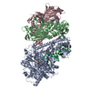
EMDB-51330:
Structure of WT human mitochondrial DNA polymerase gamma
Method: single particle / : Valenzuela S, Falkenberg M
PDB-9ggb:
Structure of the G848S mutant of human mitochondrial DNA polymerase gamma in complex with PZL-A
Method: single particle / : Valenzuela S, Falkenberg M
PDB-9ggc:
Structure of the G848S mutant of human mitochondrial DNA polymerase gamma
Method: single particle / : Valenzuela S, Falkenberg M
PDB-9ggd:
Structure of the A467T mutant of human mitochondrial DNA polymerase gamma in complex with PZL-A
Method: single particle / : Valenzuela S, Falkenberg M
PDB-9gge:
Structure of the A467T mutant of human mitochondrial DNA polymerase gamma
Method: single particle / : Valenzuela S, Falkenberg M
PDB-9ggf:
Structure of WT human mitochondrial DNA polymerase gamma
Method: single particle / : Valenzuela S, Falkenberg M
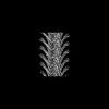
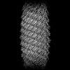
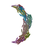
EMDB-50748:
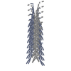
Asgard ESCRT-IIIB filament structure
Method: helical / : Chaaban S, Souza DP, Baum B
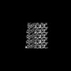
EMDB-50749:
Asgard ESCRT-IIIB membrane-bound protofilament structure
Method: helical / : Chaaban S, Souza DP, Baum B
PDB-9ftm:
Asgard ESCRT-IIIB membrane-bound protofilament structure
Method: helical / : Chaaban S, Souza DP, Baum B
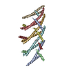
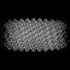
EMDB-18318:
Cryo-EM structure of Vipp1 helical filament with lattice 1 (Vipp1_L1)
Method: helical / : Naskar S, Low HH
EMDB-18321:
Cryo-EM structure of Vipp1-deltaH6_aa1-219 helical filament with lattice 2 (Vipp1-deltaH6_L2)
Method: helical / : Naskar S, Low HH
PDB-8qbr:
Cryo-EM structure of Vipp1 helical filament with lattice 1 (Vipp1_L1)
Method: helical / : Naskar S, Low HH
PDB-8qbv:
Cryo-EM structure of Vipp1-deltaH6_aa1-219 helical filament with lattice 2 (Vipp1-deltaH6_L2)
Method: helical / : Naskar S, Low HH
EMDB-18319:
Cryo-EM structure of Vipp1-F197K/L200K helical filament with lattice 1 (Vipp1-F197K/L200K_L1)
Method: helical / : Naskar S, Low HH
EMDB-18322:
Cryo-EM structure of Vipp1-deltaH6_aa1-219 helical filament with lattice 3 (Vipp1-deltaH6_L3)
Method: helical / : Naskar S, Low HH
PDB-8qbs:
Cryo-EM structure of Vipp1-F197K/L200K helical filament with lattice 1 (Vipp1-F197K/L200K_L1)
Method: helical / : Naskar S, Low HH
PDB-8qbw:
Cryo-EM structure of Vipp1-deltaH6_aa1-219 helical filament with lattice 3 (Vipp1-deltaH6_L3)
Method: helical / : Naskar S, Low HH

EMDB-50592:
SOLIST native mouse heart muscle tomogram #3
Method: electron tomography / : Erdmann PS, Nguyen HTD, Perone G, Klena N, Vazzana R, Kaluthantrige Don F, Silva M, Sorrentino S, Swuec P, Leroux F, Kalebic N, Coscia F

EMDB-50603:
SOLIST cryo-tomogram of native mouse corpus callosum #1
Method: electron tomography / : Erdmann PS, Nguyen HTD, Perone G, Klena N, Vazzana R, Kaluthantrige Don F, Silva M, Sorrentino S, Swuec P, Leroux F, Kalebic N, Coscia F

EMDB-50604:
SOLIST cryo-tomogram of native mouse corpus callosum #2
Method: electron tomography / : Erdmann PS, Nguyen HTD, Perone G, Klena N, Vazzana R, Kaluthantrige Don F, Silva M, Sorrentino S, Swuec P, Leroux F, Kalebic N, Coscia F

EMDB-50605:
native mouse liver solist tomogram #1
Method: electron tomography / : Erdmann PS, Nguyen HTD, Perone G, Klena N, Vazzana R, Kaluthantrige Don F, Silva M, Sorrentino S, Swuec P, Leroux F, Kalebic N, Coscia F

EMDB-50606:
native mouse liver solist tomogram #2
Method: electron tomography / : Erdmann PS, Nguyen HTD, Perone G, Klena N, Vazzana R, Kaluthantrige Don F, Silva M, Sorrentino S, Swuec P, Leroux F, Kalebic N, Coscia F

EMDB-50607:
SOLIST cryo-tomogram of human brain organoid #1
Method: electron tomography / : Erdmann PS, Nguyen HTD, Perone G, Klena N, Vazzana R, Kaluthantrige Don F, Silva M, Sorrentino S, Swuec P, Leroux F, Kalebic N, Coscia F

EMDB-50608:
SOLIST cryo-tomogram of human brain organoid #2
Method: electron tomography / : Erdmann PS, Nguyen HTD, Perone G, Klena N, Vazzana R, Kaluthantrige Don F, Silva M, Sorrentino S, Swuec P, Leroux F, Kalebic N, Coscia F

EMDB-50620:
S. cerevisiae ribosome from SOLIST lamellas
Method: subtomogram averaging / : Erdmann PS, Nguyen HTD, Perone G, Klena N, Vazzana R, Kaluthantrige Don F, Silva M, Sorrentino S, Swuec P, Leroux F, Kalebic N, Coscia F

EMDB-50630:
mus musculus ribosome from SOLIST lamellas
Method: subtomogram averaging / : Erdmann PS, Nguyen HTD, Perone G, Klena N, Vazzana R, Kaluthantrige Don F, Silva M, Sorrentino S, Swuec P, Leroux F, Kalebic N, Coscia F

EMDB-50646:
mus musculus heart thin filament from SOLIST lamellas
Method: subtomogram averaging / : Erdmann PS, Nguyen HTD, Perone G, Klena N, Vazzana R, Kaluthantrige Don F, Silva M, Sorrentino S, Swuec P, Leroux F, Kalebic N, Coscia F

EMDB-50655:
mus musculus heart thin filament with myosin heads from SOLIST lamellas
Method: subtomogram averaging / : Erdmann PS, Nguyen HTD, Perone G, Klena N, Vazzana R, Kaluthantrige Don F, Silva M, Sorrentino S, Swuec P, Leroux F, Kalebic N, Coscia F
EMDB-19822:
Helical reconstruction of yeast eisosome protein Pil1 bound to membrane composed of lipid mixture +PIP2/+bromosterol (DOPC, DOPE, DOPS, bromo-ergosterol, PI(4,5)P2 35:20:20:15:10)
Method: helical / : Kefauver JM, Zou L, Desfosses A, Loewith RJ
EMDB-18307:
Native eisosome lattice bound to plasma membrane microdomain
Method: single particle / : Kefauver JM, Zou L, Loewith RJ, Desfosses A
EMDB-18308:
Helical reconstruction of yeast eisosome protein Pil1 bound to membrane composed of lipid mixture -PIP2/+sterol (DOPC, DOPE, DOPS, cholesterol 30:20:20:30)
Method: helical / : Kefauver JM, Zou L, Desfosses A, Loewith RJ
EMDB-18309:
Helical reconstruction of yeast eisosome protein Pil1 bound to membrane composed of lipid mixture +PIP2/-sterol (DOPC, DOPE, DOPS, PI(4,5)P2 50:20:20:10)
Method: helical / : Kefauver JM, Zou L, Desfosses A, Loewith RJ
EMDB-18310:
Helical reconstruction of yeast eisosome protein Pil1 bound to membrane composed of lipid mixture +PIP2/+sterol (DOPC, DOPE, DOPS, cholesterol, PI(4,5)P2 35:20:20:15:10)
Method: helical / : Kefauver JM, Zou L, Desfosses A, Loewith RJ
EMDB-18311:
Compact state - Native eisosome lattice bound to plasma membrane microdomain
Method: single particle / : Kefauver JM, Zou L, Desfosses A, Loewith RJ
EMDB-18312:
Stretched state - Native eisosome lattice bound to plasma membrane microdomain
Method: single particle / : Kefauver JM, Zou L, Desfosses A, Loewith RJ
PDB-8qb7:
Pil1 in native eisosome lattice bound to plasma membrane microdomain
Method: single particle / : Kefauver JM, Zou L, Loewith RJ, Desfosses A
PDB-8qb8:
Lsp1 in native eisosome lattice bound to plasma membrane microdomain
Method: single particle / : Kefauver JM, Zou L, Loewith RJ, Desfosses A
PDB-8qb9:
Helical reconstruction of yeast eisosome protein Pil1 bound to membrane composed of lipid mixture -PIP2/+sterol (DOPC, DOPE, DOPS, cholesterol 30:20:20:30)
Method: helical / : Kefauver JM, Zou L, Desfosses A, Loewith RJ
PDB-8qbb:
Helical reconstruction of yeast eisosome protein Pil1 bound to membrane composed of lipid mixture +PIP2/-sterol (DOPC, DOPE, DOPS, PI(4,5)P2 50:20:20:10)
Method: helical / : Kefauver JM, Zou L, Desfosses A, Loewith RJ
PDB-8qbd:
Helical reconstruction of yeast eisosome protein Pil1 bound to membrane composed of lipid mixture +PIP2/+sterol (DOPC, DOPE, DOPS, cholesterol, PI(4,5)P2 35:20:20:15:10)
Method: helical / : Kefauver JM, Zou L, Desfosses A, Loewith RJ
PDB-8qbe:
Compact state - Pil1 in native eisosome lattice bound to plasma membrane microdomain
Method: single particle / : Kefauver JM, Zou L, Desfosses A, Loewith RJ
PDB-8qbf:
Compact state - Pil1 dimer with lipid headgroups fitted in native eisosome lattice bound to plasma membrane microdomain
Method: single particle / : Kefauver JM, Zou L, Desfosses A, Loewith RJ
PDB-8qbg:
Stretched state - Pil1 in native eisosome lattice bound to plasma membrane microdomain
Method: single particle / : Kefauver JM, Zou L, Desfosses A, Loewith RJ
Pages:
 wwPDB to switch to version 3 of the EMDB data model
