 Movie
Movie Controller

[English] 日本語
 Yorodumi
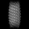
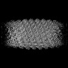
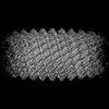
- PDB-8qbr: Cryo-EM structure of Vipp1 helical filament with lattice 1 (Vipp1_L1) -
+ Open data

- Basic information
| Entry | Database: PDB / ID: 8qbr | ||||||
|---|---|---|---|---|---|---|---|
| Title | Cryo-EM structure of Vipp1 helical filament with lattice 1 (Vipp1_L1) | ||||||
 Components Components | Membrane-associated protein Vipp1 | ||||||
 Keywords Keywords | LIPID BINDING PROTEIN / Vipp1/IM30/ESCRT-III / Membrane remodeling / Cryoelectron microscopy / Helical filament structure | ||||||
| Function / homology | PspA/IM30 / PspA/IM30 family / lipid binding / plasma membrane / Membrane-associated protein Vipp1 Function and homology information Function and homology information | ||||||
| Biological species |  Nostoc punctiforme (bacteria) Nostoc punctiforme (bacteria) | ||||||
| Method | ELECTRON MICROSCOPY / helical reconstruction / cryo EM / Resolution: 3.74 Å | ||||||
 Authors Authors | Naskar, S. / Low, H.H. | ||||||
| Funding support |  United Kingdom, 1items United Kingdom, 1items
| ||||||
 Citation Citation | Journal: Nat Struct Mol Biol / Year: 2025 Title: Mechanism for Vipp1 spiral formation, ring biogenesis, and membrane repair. Authors: Souvik Naskar / Andrea Merino / Javier Espadas / Jayanti Singh / Aurelien Roux / Adai Colom / Harry H Low /   Abstract: The ESCRT-III-like protein Vipp1 couples filament polymerization with membrane remodeling. It assembles planar sheets as well as 3D rings and helical polymers, all implicated in mitigating plastid- ...The ESCRT-III-like protein Vipp1 couples filament polymerization with membrane remodeling. It assembles planar sheets as well as 3D rings and helical polymers, all implicated in mitigating plastid-associated membrane stress. The architecture of Vipp1 planar sheets and helical polymers remains unknown, as do the geometric changes required to transition between polymeric forms. Here we show how cyanobacterial Vipp1 assembles into morphologically-related sheets and spirals on membranes in vitro. The spirals converge to form a central ring similar to those described in membrane budding. Cryo-EM structures of helical filaments reveal a close geometric relationship between Vipp1 helical and planar lattices. Moreover, the helical structures reveal how filaments twist-a process required for Vipp1, and likely other ESCRT-III filaments, to transition between planar and 3D architectures. Overall, our results provide a molecular model for Vipp1 ring biogenesis and a mechanism for Vipp1 membrane stabilization and repair, with implications for other ESCRT-III systems. | ||||||
| History |
|
- Structure visualization
| Structure viewer | Molecule: MolmilJmol/JSmol |
|---|
- Downloads & links
-Download
| PDBx/mmCIF format | 8qbr.cif.gz | 83.4 KB | Display | PDBx/mmCIF format |
|---|---|---|---|---|
| PDB format | pdb8qbr.ent.gz | 65 KB | Display | PDB format |
| PDBx/mmJSON format | 8qbr.json.gz | Tree view | PDBx/mmJSON format | |
| Others |  Other downloads Other downloads |
-Validation report
| Arichive directory | https://data.pdbj.org/pub/pdb/validation_reports/qb/8qbrftp://data.pdbj.org/pub/pdb/validation_reports/qb/8qbr | HTTPS FTP |
|---|
-Related structure data
| Related structure data | 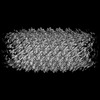 18318MC  8qbsC  8qbvC  8qbwC M: map data used to model this data C: citing same article ( |
|---|---|
| Similar structure data |
-Links
 PDBj
PDBj- Assembly
| Deposited unit | 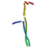
|
|---|---|
| 1 | x 95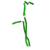
|
-Components
| #1: Protein | Mass: 23962.100 Da / Num. of mol.: 1 Source method: isolated from a genetically manipulated source Source: (gene. exp.) Nostoc punctiforme (bacteria) / Gene: vipp1, Npun_R3963 / Production host: |
|---|---|
| Has protein modification | N |
-Experimental details
-Experiment
| Experiment | Method: ELECTRON MICROSCOPY |
|---|---|
| EM experiment | Aggregation state: FILAMENT / 3D reconstruction method: helical reconstruction |
- Sample preparation
| Component | Name: Vipp1_L1 / Type: COMPLEX / Entity ID: all / Source: RECOMBINANT |
|---|---|
| Molecular weight | Experimental value: NO |
| Source (natural) | Organism: Nostoc punctiforme (bacteria) |
| Source (recombinant) | Organism: |
| Buffer solution | pH: 8.4 |
| Specimen | Conc.: 1.7 mg/ml / Embedding applied: NO / Shadowing applied: NO / Staining applied: NO / Vitrification applied: YES |
| Specimen support | Grid material: COPPER / Grid mesh size: 300 divisions/in. / Grid type: Quantifoil R2/2 |
| Vitrification | Instrument: FEI VITROBOT MARK IV / Cryogen name: ETHANE / Humidity: 100 % / Chamber temperature: 283.15 K |
- Electron microscopy imaging
| Experimental equipment |  Model: Titan Krios / Image courtesy: FEI Company |
|---|---|
| Microscopy | Model: FEI TITAN KRIOS |
| Electron gun | Electron source:  FIELD EMISSION GUN / Accelerating voltage: 300 kV / Illumination mode: FLOOD BEAM FIELD EMISSION GUN / Accelerating voltage: 300 kV / Illumination mode: FLOOD BEAM |
| Electron lens | Mode: BRIGHT FIELD / Nominal defocus max: 2500 nm / Nominal defocus min: 750 nm |
| Image recording | Electron dose: 1.02 e/Å2 / Detector mode: SUPER-RESOLUTION / Film or detector model: GATAN K2 SUMMIT (4k x 4k) |
- Processing
| EM software |
| |||||||||||||||||||||||||||||||||||
|---|---|---|---|---|---|---|---|---|---|---|---|---|---|---|---|---|---|---|---|---|---|---|---|---|---|---|---|---|---|---|---|---|---|---|---|---|
| CTF correction | Type: PHASE FLIPPING AND AMPLITUDE CORRECTION | |||||||||||||||||||||||||||||||||||
| Helical symmerty | Angular rotation/subunit: -75.860068 ° / Axial rise/subunit: 2.372081 Å / Axial symmetry: C1 | |||||||||||||||||||||||||||||||||||
| 3D reconstruction | Resolution: 3.74 Å / Resolution method: FSC 0.143 CUT-OFF / Num. of particles: 43480 / Symmetry type: HELICAL | |||||||||||||||||||||||||||||||||||
| Atomic model building | B value: 91.05 / Space: REAL |
