 ムービー
ムービー コントローラー
 構造ビューア
 EMN検索について


-検索条件
-検索結果
検索 (著者・登録者: pinto & a)の結果184件中、1から50件目までを表示しています

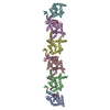
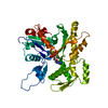
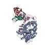
EMDB-44552: 
Septin Hexameric Complex SEPT2/SEPT6/SEPT7 of Ciona intestinalis by Cryo-EM

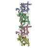
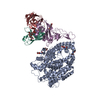
EMDB-44555:
Septin Tetrameric Complex SEPT7/SEPT9 of Ciona intestinalis by Cryo-EM
PDB-9bht:
Septin Hexameric Complex SEPT2/SEPT6/SEPT7 of Ciona intestinalis by Cryo-EM
PDB-9bhw:
Septin Tetrameric Complex SEPT7/SEPT9 of Ciona intestinalis by Cryo-EM

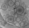
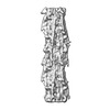
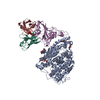
EMDB-16813:
Tomogram of GBP1 coatomers assembled on brain polar lipid-derived small unilamellar vesicles.
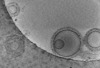
EMDB-16814:
Tomogram of GBP1 coatomers assembled on brain polar lipid-derived small unilamellar vesicles.
EMDB-16815:
Tomogram of GBP1 coatomers assembled on brain polar lipid-derived small unilamellar vesicles.
EMDB-16794:
Cryo-EM structure of the human GBP1 dimer bound to GDP-AlF3

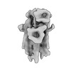
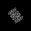
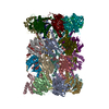
EMDB-42681:
The structure of the native cardiac thin filament troponin core in Ca2+-free state from the upper strand

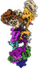
EMDB-42682:
The structure of the native cardiac thin filament troponin core in Ca2+-free tilted state from the upper strand
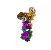
EMDB-42683:
The structure of the native cardiac thin filament troponin core in Ca2+-free rotated state from the upper strand

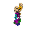
EMDB-42800:
The structure of the native cardiac thin filament troponin core in Ca2+-free state from the lower strand

EMDB-42833:
The structure of the native cardiac thin filament troponin core in Ca2+-free rotated state from the lower strand

EMDB-42835:
The structure of the native cardiac thin filament troponin core in Ca2+-free tilted state from the lower strand

EMDB-42846:
The structure of the native cardiac thin filament troponin core in Ca2+-bound fully activated state from the upper strand

EMDB-42847:
The structure of the native cardiac thin filament troponin core in Ca2+-bound partially activated state from the upper strand

EMDB-42849:
The structure of the native cardiac thin filament troponin core in Ca2+-bound fully activated state 1 from the lower strand

EMDB-42856:
The structure of the native cardiac thin filament troponin core in Ca2+-bound fully activated state 2 from the lower strand

EMDB-42858:
The structure of the native cardiac thin filament troponin core in Ca2+-bound partially activated state from the lower strand

EMDB-42874:
The structure of the native cardiac thin filament troponin core in Ca2+-free state from the upper strand activated by the C1-domain of cardiac myosin binding protein C

PDB-8uww:
The structure of the native cardiac thin filament troponin core in Ca2+-free state from the upper strand

PDB-8uwx:
The structure of the native cardiac thin filament troponin core in Ca2+-free tilted state from the upper strand
PDB-8uwy:
The structure of the native cardiac thin filament troponin core in Ca2+-free rotated state from the upper strand

PDB-8uyd:
The structure of the native cardiac thin filament troponin core in Ca2+-free state from the lower strand

PDB-8uz5:
The structure of the native cardiac thin filament troponin core in Ca2+-free rotated state from the lower strand

PDB-8uz6:
The structure of the native cardiac thin filament troponin core in Ca2+-free tilted state from the lower strand

PDB-8uzx:
The structure of the native cardiac thin filament troponin core in Ca2+-bound fully activated state from the upper strand

PDB-8uzy:
The structure of the native cardiac thin filament troponin core in Ca2+-bound partially activated state from the upper strand

PDB-8v01:
The structure of the native cardiac thin filament troponin core in Ca2+-bound fully activated state 1 from the lower strand

PDB-8v0i:
The structure of the native cardiac thin filament troponin core in Ca2+-bound fully activated state 2 from the lower strand

PDB-8v0k:
The structure of the native cardiac thin filament troponin core in Ca2+-bound partially activated state from the lower strand

PDB-8v0y:
The structure of the native cardiac thin filament troponin core in Ca2+-free state from the upper strand activated by the C1-domain of cardiac myosin binding protein C
EMDB-16776:
Human arginylated beta-actin
PDB-8cog:
Human arginylated beta-actin
EMDB-40468:
In situ human cardiac thick filament in the relaxed state
EMDB-40471:
Human cardiac interacting heads motif (IHM-C) in complete form
EMDB-40475:
Human cardiac interacting heads motif (IHM-C) in semi form
EMDB-40476:
Human cardiac interacting heads motif (IHM-S)
EMDB-40478:
Human cardiac interacting heads motif (IHM-D) in semi form

EMDB-29530:
SARS-CoV-2 XBB.1 spike RBD bound to the human ACE2 ectodomain and the S309 neutralizing antibody Fab fragment
EMDB-29531:
SARS-CoV-2 BQ.1.1 spike RBD bound to the human ACE2 ectodomain and the S309 neutralizing antibody Fab fragment
EMDB-40240:
SARS-CoV-2 BN.1 spike RBD bound to the human ACE2 ectodomain and the S309 neutralizing antibody Fab fragment
PDB-8fxb:
SARS-CoV-2 XBB.1 spike RBD bound to the human ACE2 ectodomain and the S309 neutralizing antibody Fab fragment
PDB-8fxc:
SARS-CoV-2 BQ.1.1 spike RBD bound to the human ACE2 ectodomain and the S309 neutralizing antibody Fab fragment
PDB-8s9g:
SARS-CoV-2 BN.1 spike RBD bound to the human ACE2 ectodomain and the S309 neutralizing antibody Fab fragment
EMDB-16963:
Leishmania tarentolae proteasome 20S subunit in complex with 1-Benzyl-N-(3-(cyclopropylcarbamoyl)phenyl)-6-oxo-1,6-dihydropyridazine-3-carboxamide
PDB-8olu:
Leishmania tarentolae proteasome 20S subunit in complex with 1-Benzyl-N-(3-(cyclopropylcarbamoyl)phenyl)-6-oxo-1,6-dihydropyridazine-3-carboxamide
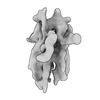
EMDB-40245:
Human ER membrane protein complex (EMC) in GDN, 9-subunit map
EMDB-40246:
Human ER membrane protein complex (EMC) in GDN, Consensus map
EMDB-40247:
Human ER membrane protein complex (EMC) in GDN, 8-subunit map
ページ:
 wwPDBはEMDBデータモデルのバージョン3へ移行します
