 Movie
Movie Controller


+ Open data

- Basic information
| Entry | Database: PDB / ID: 8cog | ||||||||||||||||||||||||
|---|---|---|---|---|---|---|---|---|---|---|---|---|---|---|---|---|---|---|---|---|---|---|---|---|---|
| Title | Human arginylated beta-actin | ||||||||||||||||||||||||
 Components Components | Actin, cytoplasmic 1, N-terminally processed | ||||||||||||||||||||||||
 Keywords Keywords | CONTRACTILE PROTEIN / actin / methylated / filament | ||||||||||||||||||||||||
| Function / homology |  Function and homology information Function and homology informationpositive regulation of norepinephrine uptake / cellular response to cytochalasin B / Formation of the embryonic stem cell BAF (esBAF) complex / bBAF complex / npBAF complex / brahma complex / nBAF complex / Formation of the canonical BAF (cBAF) complex / regulation of transepithelial transport / Formation of neuronal progenitor and neuronal BAF (npBAF and nBAF) ...positive regulation of norepinephrine uptake / cellular response to cytochalasin B / Formation of the embryonic stem cell BAF (esBAF) complex / bBAF complex / npBAF complex / brahma complex / nBAF complex / Formation of the canonical BAF (cBAF) complex / regulation of transepithelial transport / Formation of neuronal progenitor and neuronal BAF (npBAF and nBAF) / morphogenesis of a polarized epithelium / Formation of the polybromo-BAF (pBAF) complex / structural constituent of postsynaptic actin cytoskeleton / Formation of annular gap junctions / Formation of the dystrophin-glycoprotein complex (DGC) / Gap junction degradation / Formation of the non-canonical BAF (ncBAF) complex / GBAF complex / protein localization to adherens junction / regulation of G0 to G1 transition / Cell-extracellular matrix interactions / dense body / Folding of actin by CCT/TriC / Tat protein binding / postsynaptic actin cytoskeleton / RSC-type complex / Regulation of CDH1 Function / regulation of double-strand break repair / regulation of nucleotide-excision repair / Prefoldin mediated transfer of substrate to CCT/TriC / Adherens junctions interactions / adherens junction assembly / RHOF GTPase cycle / apical protein localization / Sensory processing of sound by outer hair cells of the cochlea / SWI/SNF complex / regulation of mitotic metaphase/anaphase transition / tight junction / Sensory processing of sound by inner hair cells of the cochlea / Interaction between L1 and Ankyrins / positive regulation of T cell differentiation / apical junction complex / positive regulation of double-strand break repair / maintenance of blood-brain barrier / regulation of norepinephrine uptake / positive regulation of stem cell population maintenance / transporter regulator activity / NuA4 histone acetyltransferase complex / Recycling pathway of L1 / Regulation of MITF-M-dependent genes involved in pigmentation / cortical cytoskeleton / establishment or maintenance of cell polarity / nitric-oxide synthase binding / brush border / regulation of G1/S transition of mitotic cell cycle / EPH-ephrin mediated repulsion of cells / negative regulation of cell differentiation / regulation of synaptic vesicle endocytosis / positive regulation of myoblast differentiation / RHO GTPases Activate WASPs and WAVEs / kinesin binding / regulation of protein localization to plasma membrane / RHO GTPases activate IQGAPs / positive regulation of double-strand break repair via homologous recombination / EPHB-mediated forward signaling / cytoskeleton organization / axonogenesis / substantia nigra development / calyx of Held / nitric-oxide synthase regulator activity / FCGR3A-mediated phagocytosis / Translocation of SLC2A4 (GLUT4) to the plasma membrane / actin filament / adherens junction / positive regulation of cell differentiation / Regulation of endogenous retroelements by Piwi-interacting RNAs (piRNAs) / cell motility / RHO GTPases Activate Formins / Signaling by high-kinase activity BRAF mutants / MAP2K and MAPK activation / Regulation of actin dynamics for phagocytic cup formation / structural constituent of cytoskeleton / kinetochore / B-WICH complex positively regulates rRNA expression / VEGFA-VEGFR2 Pathway / platelet aggregation / DNA Damage Recognition in GG-NER / Hydrolases; Acting on acid anhydrides; Acting on acid anhydrides to facilitate cellular and subcellular movement / tau protein binding / Schaffer collateral - CA1 synapse / nuclear matrix / Signaling by RAF1 mutants / cytoplasmic ribonucleoprotein granule / Signaling by moderate kinase activity BRAF mutants / Paradoxical activation of RAF signaling by kinase inactive BRAF / Signaling downstream of RAS mutants / cell-cell junction / Signaling by BRAF and RAF1 fusions / UCH proteinases / nucleosome Similarity search - Function | ||||||||||||||||||||||||
| Biological species |  Homo sapiens (human) Homo sapiens (human) | ||||||||||||||||||||||||
| Method | ELECTRON MICROSCOPY / single particle reconstruction / cryo EM / Resolution: 3.499 Å | ||||||||||||||||||||||||
 Authors Authors | Pinto, C.S. / Bakker, S.E. / Suchenko, A. / Hussain, H. / Hatano, T. / Sampath, K. / Chinthalapudi, K. / Mishima, M. / Balasubramanian, M. | ||||||||||||||||||||||||
| Funding support |  United Kingdom, European Union, 4items United Kingdom, European Union, 4items
| ||||||||||||||||||||||||
 Citation Citation | Journal: J Cell Biol / Year: 2026 Title: Actin arginylation alters myosin engagement and F-actin patterning despite structural conservation. Authors: Clyde Savio Pinto / Saskia E Bakker / Andrejus Suchenko / Isabella M Kolodny / Hamdi Hussain / Tomoyuki Hatano / Karuna Sampath / Krishna Chinthalapudi / Sarah M Heissler / Masanori Mishima ...Authors: Clyde Savio Pinto / Saskia E Bakker / Andrejus Suchenko / Isabella M Kolodny / Hamdi Hussain / Tomoyuki Hatano / Karuna Sampath / Krishna Chinthalapudi / Sarah M Heissler / Masanori Mishima / Mohan Balasubramanian /  Abstract: Actin is a conserved protein with crucial roles in cell polarity, division, and muscle contraction. Its function is regulated in part by posttranslational modifications, one of which is N-terminal ...Actin is a conserved protein with crucial roles in cell polarity, division, and muscle contraction. Its function is regulated in part by posttranslational modifications, one of which is N-terminal arginylation. What is the structure of arginylated-β-actin (R-β-actin), and how does it regulate F-actin function? Here we report the 3.6 Å structures of ADP-R-β-actin filaments, which are nearly identical to that of non-arginylated F-actin. In vitro assays reveal that the interaction between myosin-II and actin is altered upon actin arginylation, characterized by frequent detachment of R-actin filaments from myosin-II. In vivo, replacement of the only actin gene in Schizosaccharomyces pombe with a synthetic gene encoding R-Sp-actin reduces Arp2/3-based actin patches while thickening formin-induced actin cables. Consistent with defective interactions between myosin-II and R-actin filaments, assembly and constriction of the cytokinetic actomyosin ring are perturbed in R-Sp-actin cells. Thus, despite structural similarity of arginylated and non-arginylated actin filaments, actin arginylation affects F-actin assortment into distinct subcellular structures and its interaction with myosin-II. | ||||||||||||||||||||||||
| History |
|
- Structure visualization
| Structure viewer | Molecule: MolmilJmol/JSmol |
|---|
- Downloads & links
-Download
| PDBx/mmCIF format | 8cog.cif.gz | 90 KB | Display | PDBx/mmCIF format |
|---|---|---|---|---|
| PDB format | pdb8cog.ent.gz | 63.8 KB | Display | PDB format |
| PDBx/mmJSON format | 8cog.json.gz | Tree view | PDBx/mmJSON format | |
| Others |  Other downloads Other downloads |
-Validation report
| Arichive directory | https://data.pdbj.org/pub/pdb/validation_reports/co/8cogftp://data.pdbj.org/pub/pdb/validation_reports/co/8cog | HTTPS FTP |
|---|
-Related structure data
| Related structure data |  16776MC M: map data used to model this data C: citing same article ( |
|---|---|
| Similar structure data |
-Links
 PDBj
PDBj


- Assembly
| Deposited unit | 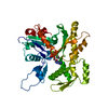
|
|---|---|
| 1 |
|
-Components
| #1: Protein | Mass: 41706.590 Da / Num. of mol.: 1 Source method: isolated from a genetically manipulated source Source: (gene. exp.) Homo sapiens (human) / Gene: ACTB / Production host:  Komagataella pastoris (fungus) / References: UniProt: P60709 Komagataella pastoris (fungus) / References: UniProt: P60709 |
|---|---|
| #2: Chemical | ChemComp-ADP /   Mass: 427.201 Da / Num. of mol.: 1 / Source method: obtained synthetically / Formula: C10H15N5O10P2 / Comment: ADP, energy-carrying molecule*YM Mass: 427.201 Da / Num. of mol.: 1 / Source method: obtained synthetically / Formula: C10H15N5O10P2 / Comment: ADP, energy-carrying molecule*YM |
| #3: Chemical | ChemComp-MG /   Mass: 24.305 Da / Num. of mol.: 1 / Source method: obtained synthetically / Formula: Mg Mass: 24.305 Da / Num. of mol.: 1 / Source method: obtained synthetically / Formula: Mg |
| Has ligand of interest | N |
| Has protein modification | Y |
-Experimental details
-Experiment
| Experiment | Method: ELECTRON MICROSCOPY |
|---|---|
| EM experiment | Aggregation state: HELICAL ARRAY / 3D reconstruction method: single particle reconstruction |
- Sample preparation
| Component | Name: Polymerised arginylated actin with bound ADP / Type: COMPLEX / Entity ID: #1 / Source: RECOMBINANT |
|---|---|
| Source (natural) | Organism: Homo sapiens (human) |
| Source (recombinant) | Organism: Komagataella pastoris (fungus) |
| Buffer solution | pH: 7.4 Details: Actin for EM was polymerized by mixing, 20 ul 20 uM G-actin, 8 ul 10x MKE and 52 ul 5 mM HEPES-KOH pH 7.4 containing 0.2 mM ATP and 0.5 mM DTT and incubating at RT for 1 hour. |
| Specimen | Embedding applied: NO / Shadowing applied: NO / Staining applied: NO / Vitrification applied: YES |
| Specimen support | Grid material: COPPER / Grid mesh size: 200 divisions/in. / Grid type: Quantifoil R3.5/1 |
| Vitrification | Instrument: LEICA EM GP / Cryogen name: ETHANE-PROPANE / Humidity: 95 % / Chamber temperature: 277 K |
- Electron microscopy imaging
| Experimental equipment |  Model: Titan Krios / Image courtesy: FEI Company |
|---|---|
| Microscopy | Model: FEI TITAN KRIOS |
| Electron gun | Electron source:  FIELD EMISSION GUN / Accelerating voltage: 300 kV / Illumination mode: FLOOD BEAM FIELD EMISSION GUN / Accelerating voltage: 300 kV / Illumination mode: FLOOD BEAM |
| Electron lens | Mode: BRIGHT FIELD / Nominal magnification: 75000 X / Nominal defocus max: 1500 nm / Nominal defocus min: 700 nm / Cs: 2.7 mm / C2 aperture diameter: 50 µm |
| Specimen holder | Cryogen: NITROGEN / Specimen holder model: FEI TITAN KRIOS AUTOGRID HOLDER |
| Image recording | Average exposure time: 60 sec. / Electron dose: 42 e/Å2 / Detector mode: COUNTING / Film or detector model: GATAN K2 SUMMIT (4k x 4k) / Num. of grids imaged: 1 |
- Processing
| Software | Name: REFMAC / Version: 5.8.0267 / Classification: refinement / Contact author: Garib N. Murshudov / Contact author email: garib[at]mrc-lmb.cam.ac.uk / Date: 2020-24-08 Description: (un)restrained refinement or idealisation of macromolecular structures | ||||||||||||||||||||||||||||||||||||||||||||||||||||||||||||||||||||||||||||||||||||||||||||||||||||||||||
|---|---|---|---|---|---|---|---|---|---|---|---|---|---|---|---|---|---|---|---|---|---|---|---|---|---|---|---|---|---|---|---|---|---|---|---|---|---|---|---|---|---|---|---|---|---|---|---|---|---|---|---|---|---|---|---|---|---|---|---|---|---|---|---|---|---|---|---|---|---|---|---|---|---|---|---|---|---|---|---|---|---|---|---|---|---|---|---|---|---|---|---|---|---|---|---|---|---|---|---|---|---|---|---|---|---|---|---|
| EM software |
| ||||||||||||||||||||||||||||||||||||||||||||||||||||||||||||||||||||||||||||||||||||||||||||||||||||||||||
| CTF correction | Type: PHASE FLIPPING AND AMPLITUDE CORRECTION | ||||||||||||||||||||||||||||||||||||||||||||||||||||||||||||||||||||||||||||||||||||||||||||||||||||||||||
| 3D reconstruction | Resolution: 3.499 Å / Resolution method: FSC 0.143 CUT-OFF / Num. of particles: 42701 / Algorithm: FOURIER SPACE Details: RELION 3.09 was used throughout using helical reconstruction. Symmetry type: POINT | ||||||||||||||||||||||||||||||||||||||||||||||||||||||||||||||||||||||||||||||||||||||||||||||||||||||||||
| Refinement | Resolution: 3.499→3.499 Å / Cor.coef. Fo:Fc: 0.806 / WRfactor Rwork: 0.36 / SU B: 20.983 / SU ML: 0.319 / Average fsc overall: 0.8232 / Average fsc work: 0.8232 / ESU R: 0.508 Details: Hydrogens have been added in their riding positions
| ||||||||||||||||||||||||||||||||||||||||||||||||||||||||||||||||||||||||||||||||||||||||||||||||||||||||||
| Solvent computation | Solvent model: BABINET MODEL | ||||||||||||||||||||||||||||||||||||||||||||||||||||||||||||||||||||||||||||||||||||||||||||||||||||||||||
| Displacement parameters | Biso mean: 77.447 Å2 | ||||||||||||||||||||||||||||||||||||||||||||||||||||||||||||||||||||||||||||||||||||||||||||||||||||||||||
| Refine LS restraints |
|