 Movie
Movie Controller
 Structure viewers
 About EMN search


-Search query
-Search result
Showing 1 - 50 of 74 items for (author: park & kh)

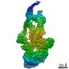
EMDB-42074: 
Representative tomogram of Enterococcus faecium WT Com15

EMDB-42086:
Representative tomogram of Enterococcus faecium SagA complementation strain

EMDB-42087:
Representative tomogram of Enterococcus faecium SagA deletion strain

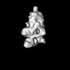
EMDB-43033:
Structure of 80alpha portal protein expressed in E. coli
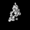
EMDB-43142:
SaPI1 mature capsid structure containing DNA

EMDB-43143:
SaPI1 mature capsid structure without DNA

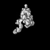
EMDB-43145:
SaPI1 portal structure in mature capsids containing DNA

EMDB-43146:
SaPI1 portal structure in mature capsids without DNA
EMDB-43147:
SaPI1 portal-capsid interface in mature capsids with DNA
PDB-8v8b:
Structure of 80alpha portal protein expressed in E. coli
PDB-8vd4:
SaPI1 mature capsid structure containing DNA
PDB-8vd5:
SaPI1 mature capsid structure without DNA
PDB-8vd8:
SaPI1 portal structure in mature capsids containing DNA
PDB-8vdc:
SaPI1 portal structure in mature capsids without DNA
PDB-8vde:
SaPI1 portal-capsid interface in mature capsids with DNA

PDB-8c5v:
Chemotaxis core signalling unit from E protein lysed E. coli cells
EMDB-27703:
Structure of RBD directed antibody DH1047 in complex with SARS-CoV-2 spike: Local refinement of RBD-Fab interace
PDB-8dtk:
Structure of RBD directed antibody DH1047 in complex with SARS-CoV-2 spike: Local refinement of RBD-Fab interace

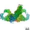
EMDB-15641:
Structure of the Native Chemotaxis Core Signalling Complex from E-gene lysed E. coli cells.
EMDB-15642:
Native Chemotaxis Core Signalling Complex from E. coli, with focused alignment on the baseplate (CheA-CheW)

EMDB-15643:
Native Chemotaxis Core Signalling Complex from E. coli, Focused alignment on ligand binding domain

EMDB-26727:
Structure of the human coronavirus CCoV-HuPn-2018 spike glycoprotein with domain 0 in the proximal conformation

EMDB-26729:
CCoV-HuPn-2018 S in the proximal conformation (local refinement of domain 0)

EMDB-26730:
Structure of the human coronavirus CCoV-HuPn-2018 spike glycoprotein with domain 0 in the swung out conformation

EMDB-26731:
CCoV-HuPn-2018 S in the swung out conformation (local refinement of domain 0)
PDB-7us6:
Structure of the human coronavirus CCoV-HuPn-2018 spike glycoprotein with domain 0 in the proximal conformation
PDB-7us9:
CCoV-HuPn-2018 S in the proximal conformation (local refinement of domain 0)
PDB-7usa:
Structure of the human coronavirus CCoV-HuPn-2018 spike glycoprotein with domain 0 in the swung out conformation
PDB-7usb:
CCoV-HuPn-2018 S in the swung out conformation (local refinement of domain 0)

EMDB-26676:
Three RBD-down state of SARS-CoV-2 D614G spike in complex with the SP1-77 neutralizing antibody Fab fragment
EMDB-26677:
Three RBD-down state of SARS-CoV-2 D614G spike in complex with the SP1-77 neutralizing antibody Fab fragment (local refinement of the RBD and Fab variable domains)
EMDB-26678:
An antibody from single human VH-rearranging mouse neutralizes all SARS-CoV-2 variants through BA.5 by inhibiting membrane fusion
EMDB-27043:
Negative stain electron microscopy single particle reconstruction of monoclonal antibody SP1-77 Fab in complex with SARS-CoV2 2P spike
EMDB-27044:
Negative stain electron microscopy single particle reconstruction of monoclonal antibody VHH7-7-53 Fab in complex with SARS-CoV2 2P spike
EMDB-27046:
Negative stain electron microscopy single particle reconstruction of monoclonal antibody VHH7-5-82 Fab in complex with SARS-CoV2 2P spike
PDB-7upw:
Three RBD-down state of SARS-CoV-2 D614G spike in complex with the SP1-77 neutralizing antibody Fab fragment
PDB-7upx:
Three RBD-down state of SARS-CoV-2 D614G spike in complex with the SP1-77 neutralizing antibody Fab fragment (local refinement of the RBD and Fab variable domains)
PDB-7upy:
An antibody from single human VH-rearranging mouse neutralizes all SARS-CoV-2 variants through BA.5 by inhibiting membrane fusion
EMDB-25105:
Structure of SARS-CoV S protein in complex with Receptor Binding Domain antibody DH1047

PDB-7sg4:
Structure of SARS-CoV S protein in complex with Receptor Binding Domain antibody DH1047
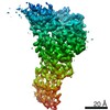
EMDB-21514:
map of the mammalian Mediator complex
PDB-6w1s:
Atomic model of the mammalian Mediator complex

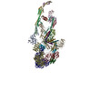
EMDB-22309:
Dimeric Immunoglobin A (dIgA)
EMDB-22310:
Secretory Immunoglobin A (SIgA)

PDB-7jg1:
Dimeric Immunoglobin A (dIgA)

PDB-7jg2:
Secretory Immunoglobin A (SIgA)
EMDB-22829:
Human Tom70 in complex with SARS CoV2 Orf9b
PDB-7kdt:
Human Tom70 in complex with SARS CoV2 Orf9b

PDB-6zet:
Crystal structure of proteinase K nanocrystals by electron diffraction with a 20 micrometre C2 condenser aperture

PDB-6zeu:
Crystal structure of proteinase K lamella by electron diffraction with a 50 micrometre C2 condenser aperture
Pages:
 wwPDB to switch to version 3 of the EMDB data model
