 Movie
Movie Controller

[English] 日本語
 Yorodumi
- PDB-6zet: Crystal structure of proteinase K nanocrystals by electron diffra... -
+ Open data

- Basic information
| Entry | Database: PDB / ID: 6zet | ||||||||||||
|---|---|---|---|---|---|---|---|---|---|---|---|---|---|
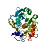
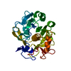
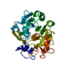
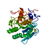
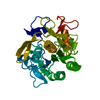
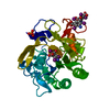
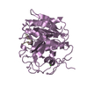
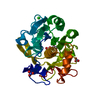
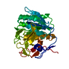
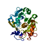
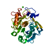
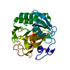
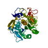
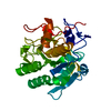
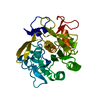
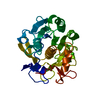
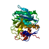
| Title | Crystal structure of proteinase K nanocrystals by electron diffraction with a 20 micrometre C2 condenser aperture | ||||||||||||
 Components Components | Proteinase K | ||||||||||||
 Keywords Keywords | HYDROLASE / Protease / Serine protease | ||||||||||||
| Function / homology |  Function and homology information Function and homology informationpeptidase K / serine-type endopeptidase activity / proteolysis / extracellular region / metal ion binding Similarity search - Function | ||||||||||||
| Biological species |  Parengyodontium album (fungus) Parengyodontium album (fungus) | ||||||||||||
| Method | ELECTRON CRYSTALLOGRAPHY / electron crystallography /  MOLECULAR REPLACEMENT / cryo EM / Resolution: 2.701 Å MOLECULAR REPLACEMENT / cryo EM / Resolution: 2.701 Å | ||||||||||||
 Authors Authors | Evans, G. / Zhang, P. / Beale, E.V. / Waterman, D.G. | ||||||||||||
| Funding support |  United Kingdom, 3items United Kingdom, 3items
| ||||||||||||
 Citation Citation | Journal: Front Mol Biosci / Year: 2020 Title: A Workflow for Protein Structure Determination From Thin Crystal Lamella by Micro-Electron Diffraction. Authors: Emma V Beale / David G Waterman / Corey Hecksel / Jason van Rooyen / James B Gilchrist / James M Parkhurst / Felix de Haas / Bart Buijsse / Gwyndaf Evans / Peijun Zhang /   Abstract: MicroED has recently emerged as a powerful method for the analysis of biological structures at atomic resolution. This technique has been largely limited to protein nanocrystals which grow either as ...MicroED has recently emerged as a powerful method for the analysis of biological structures at atomic resolution. This technique has been largely limited to protein nanocrystals which grow either as needles or plates measuring only a few hundred nanometers in thickness. Furthermore, traditional microED data processing uses established X-ray crystallography software that is not optimized for handling compound effects that are unique to electron diffraction data. Here, we present an integrated workflow for microED, from sample preparation by cryo-focused ion beam milling, through data collection with a standard Ceta-D detector, to data processing using the DIALS software suite, thus enabling routine atomic structure determination of protein crystals of any size and shape using microED. We demonstrate the effectiveness of the workflow by determining the structure of proteinase K to 2.0 Å resolution and show the advantage of using protein crystal lamellae over nanocrystals. | ||||||||||||
| History |
|
- Structure visualization
| Movie |
Movie viewer |
|---|---|
| Structure viewer | Molecule: MolmilJmol/JSmol |
- Downloads & links
-Download
| PDBx/mmCIF format | 6zet.cif.gz | 65 KB | Display | PDBx/mmCIF format |
|---|---|---|---|---|
| PDB format | pdb6zet.ent.gz | Display | PDB format | |
| PDBx/mmJSON format | 6zet.json.gz | Tree view | PDBx/mmJSON format | |
| Others |  Other downloads Other downloads |
-Validation report
| Arichive directory | https://data.pdbj.org/pub/pdb/validation_reports/ze/6zetftp://data.pdbj.org/pub/pdb/validation_reports/ze/6zet | HTTPS FTP |
|---|
-Related structure data
| Related structure data |  6zeuC  6zevC  2id8S S: Starting model for refinement C: citing same article ( |
|---|---|
| Similar structure data | |
| Experimental dataset #1 | Data reference: 10.5281/zenodo.4121985 / Data set type: diffraction image data |


-Links
 PDBj
PDBj

- Assembly
| Deposited unit | 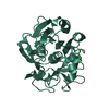
| ||||||||
|---|---|---|---|---|---|---|---|---|---|
| 1 |
| ||||||||
| Unit cell |
|
-Components
| #1: Protein | Mass: 28958.791 Da / Num. of mol.: 1 / Source method: obtained synthetically / Details: Sigma-Aldrich, P2308 / Source: (synth.) Parengyodontium album (fungus) / References: UniProt: P06873, peptidase K |
|---|---|
| #2: Chemical | ChemComp-CA /   Mass: 40.078 Da / Num. of mol.: 1 / Source method: obtained synthetically / Formula: Ca Mass: 40.078 Da / Num. of mol.: 1 / Source method: obtained synthetically / Formula: Ca |
| Has ligand of interest | N |
| Has protein modification | Y |
-Experimental details
-Experiment
| Experiment | Method: ELECTRON CRYSTALLOGRAPHY |
|---|---|
| EM experiment | Aggregation state: 3D ARRAY / 3D reconstruction method: electron crystallography |
- Sample preparation
| Component | Name: proteinase K / Type: COMPLEX / Source: MULTIPLE SOURCES |
|---|---|
| Buffer solution | pH: 7.5 |
| Buffer component | Conc.: 25 mM / Name: Tris |
| Specimen | Conc.: 50 mg/ml / Embedding applied: NO / Shadowing applied: NO / Staining applied: NO / Vitrification applied: YES |
| Specimen support | Grid type: Quantifoil R1.2/1.3 |
| Vitrification | Cryogen name: ETHANE / Humidity: 100 % / Chamber temperature: 293.15 K Details: Excess liquid was removed by blotting for 4-6s with a Vitrobot |
-Data collection
| Experimental equipment |  Model: Talos Arctica / Image courtesy: FEI Company | ||||||||||||||||||
|---|---|---|---|---|---|---|---|---|---|---|---|---|---|---|---|---|---|---|---|
| Microscopy | Model: FEI TALOS ARCTICA / Date: Apr 20, 2018 | ||||||||||||||||||
| Electron gun | Electron source: FIELD EMISSION GUN / Accelerating voltage: 200 kV / Illumination mode: FLOOD BEAM | ||||||||||||||||||
| Electron lens | Mode: DIFFRACTION / C2 aperture diameter: 20 µm | ||||||||||||||||||
| Image recording | Imaging-ID: 1 / Average exposure time: 0.85 sec. / Electron dose: 0.034 e/Å2 / Film or detector model: OTHER / Num. of grids imaged: 1 / Details: The images were recorded using a Ceta-D detector operated in rolling-shutter mode with 2x2 binning.
| ||||||||||||||||||
| Image scans | Sampling size: 28 µm / Width: 2048 / Height: 2048 | ||||||||||||||||||
| EM diffraction | Camera length: 1750 mm | ||||||||||||||||||
| EM diffraction stats | Fourier space coverage: 88.5 % / High resolution: 2.7 Å / Num. of intensities measured: 78458 / Num. of structure factors: 6274 / Phase error: 0 ° / Phase residual: 0.1 ° / Phase error rejection criteria: none / Rmerge: 0.51 / Rsym: 0.51 | ||||||||||||||||||
| Reflection shell | Resolution: 2.7→2.83 Å / Redundancy: 10.5 % / Rmerge F all: 1.313 / Rmerge(I) all: 1.275 / Rmerge(I) obs: 1.275 / Mean I/σ(I) obs: 1.8 / Num. measured obs: 8010 / Num. unique obs: 760 / CC1/2: 0.498 / Rpim(I) all: 0.533 / Rrim(I) all: 1.389 / Χ2: 0.89 / % possible all: 83.6 |
- Processing
| Software |
| |||||||||||||||||||||||||||||||||||||||||||||||||||||||||||||||||||||||||||||||||||||||||||||||||||||||||||||||||||||||||||||||||||||||||||||||||||
|---|---|---|---|---|---|---|---|---|---|---|---|---|---|---|---|---|---|---|---|---|---|---|---|---|---|---|---|---|---|---|---|---|---|---|---|---|---|---|---|---|---|---|---|---|---|---|---|---|---|---|---|---|---|---|---|---|---|---|---|---|---|---|---|---|---|---|---|---|---|---|---|---|---|---|---|---|---|---|---|---|---|---|---|---|---|---|---|---|---|---|---|---|---|---|---|---|---|---|---|---|---|---|---|---|---|---|---|---|---|---|---|---|---|---|---|---|---|---|---|---|---|---|---|---|---|---|---|---|---|---|---|---|---|---|---|---|---|---|---|---|---|---|---|---|---|---|---|---|
| EM software |
| |||||||||||||||||||||||||||||||||||||||||||||||||||||||||||||||||||||||||||||||||||||||||||||||||||||||||||||||||||||||||||||||||||||||||||||||||||
| EM 3D crystal entity | ∠α: 90 ° / ∠β: 90 ° / ∠γ: 90 ° / A: 67.3653 Å / B: 67.3653 Å / C: 106.784 Å / Space group name: P43212 / Space group num: 96 | |||||||||||||||||||||||||||||||||||||||||||||||||||||||||||||||||||||||||||||||||||||||||||||||||||||||||||||||||||||||||||||||||||||||||||||||||||
| 3D reconstruction | Resolution method: DIFFRACTION PATTERN/LAYERLINES / Symmetry type: 3D CRYSTAL | |||||||||||||||||||||||||||||||||||||||||||||||||||||||||||||||||||||||||||||||||||||||||||||||||||||||||||||||||||||||||||||||||||||||||||||||||||
| Atomic model building | Space: RECIPROCAL | |||||||||||||||||||||||||||||||||||||||||||||||||||||||||||||||||||||||||||||||||||||||||||||||||||||||||||||||||||||||||||||||||||||||||||||||||||
| Atomic model building | PDB-ID: 2ID8 Accession code: 2ID8 / Source name: PDB / Type: experimental model | |||||||||||||||||||||||||||||||||||||||||||||||||||||||||||||||||||||||||||||||||||||||||||||||||||||||||||||||||||||||||||||||||||||||||||||||||||
| Refinement | Method to determine structure: MOLECULAR REPLACEMENT Starting model: 2ID8 Resolution: 2.701→57.04 Å / Cor.coef. Fo:Fc: 0.872 / Cor.coef. Fo:Fc free: 0.824 / WRfactor Rfree: 0.239 / WRfactor Rwork: 0.204 / Average fsc free: 0.8762 / Average fsc work: 0.8959 / Cross valid method: FREE R-VALUE / ESU R Free: 0.454 Details: Hydrogens have been added in their riding positions
| |||||||||||||||||||||||||||||||||||||||||||||||||||||||||||||||||||||||||||||||||||||||||||||||||||||||||||||||||||||||||||||||||||||||||||||||||||
| Solvent computation | Ion probe radii: 0.8 Å / Shrinkage radii: 0.8 Å / VDW probe radii: 1.2 Å / Solvent model: MASK BULK SOLVENT | |||||||||||||||||||||||||||||||||||||||||||||||||||||||||||||||||||||||||||||||||||||||||||||||||||||||||||||||||||||||||||||||||||||||||||||||||||
| Displacement parameters | Biso mean: 7.281 Å2
| |||||||||||||||||||||||||||||||||||||||||||||||||||||||||||||||||||||||||||||||||||||||||||||||||||||||||||||||||||||||||||||||||||||||||||||||||||
| Refine LS restraints |
| |||||||||||||||||||||||||||||||||||||||||||||||||||||||||||||||||||||||||||||||||||||||||||||||||||||||||||||||||||||||||||||||||||||||||||||||||||
| LS refinement shell |
|
