National Institutes of Health/National Institute of General Medical Sciences (NIH/NIGMS)
5T32GM008496
United States
National Science Foundation (NSF, United States)
DMR-1548924
United States
Department of Energy (DOE, United States)
DE-FC02-02ER63421
United States
National Institutes of Health/National Institute of General Medical Sciences (NIH/NIGMS)
R35 GM128867
United States
Sao Paulo Research Foundation (FAPESP)
16/24191-8
Brazil
Sao Paulo Research Foundation (FAPESP)
17/13485-3
Brazil
Spanish Ministry of Economy and Competitiveness
BIO2015-64216-P
Spain
Spanish Ministry of Economy and Competitiveness
PGC2018-101370-B-100
Spain
Spanish Ministry of Economy and Competitiveness
MDM2014-0435-01
Spain
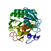
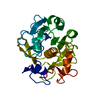
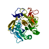
Citation
Journal: Acta Crystallogr D Struct Biol / Year: 2020 Title: Fragment-based determination of a proteinase K structure from MicroED data using ARCIMBOLDO_SHREDDER. Authors: Logan S Richards / Claudia Millán / Jennifer Miao / Michael W Martynowycz / Michael R Sawaya / Tamir Gonen / Rafael J Borges / Isabel Usón / Jose A Rodriguez / Abstract: Structure determination of novel biological macromolecules by X-ray crystallography can be facilitated by the use of small structural fragments, some of only a few residues in length, as effective ...Structure determination of novel biological macromolecules by X-ray crystallography can be facilitated by the use of small structural fragments, some of only a few residues in length, as effective search models for molecular replacement to overcome the phase problem. Independence from the need for a complete pre-existing model with sequence similarity to the crystallized molecule is the primary appeal of ARCIMBOLDO, a suite of programs which employs this ab initio algorithm for phase determination. Here, the use of ARCIMBOLDO is investigated to overcome the phase problem with the electron cryomicroscopy (cryoEM) method known as microcrystal electron diffraction (MicroED). The results support the use of the ARCIMBOLDO_SHREDDER pipeline to provide phasing solutions for a structure of proteinase K from 1.6 Å resolution data using model fragments derived from the structures of proteins sharing a sequence identity of as low as 20%. ARCIMBOLDO_SHREDDER identified the most accurate polyalanine fragments from a set of distantly related sequence homologues. Alternatively, such templates were extracted in spherical volumes and given internal degrees of freedom to refine towards the target structure. Both modes relied on the rotation function in Phaser to identify or refine fragment models and its translation function to place them. Model completion from the placed fragments proceeded through phase combination of partial solutions and/or density modification and main-chain autotracing using SHELXE. The combined set of fragments was sufficient to arrive at a solution that resembled that determined by conventional molecular replacement using the known target structure as a search model. This approach obviates the need for a single, complete and highly accurate search model when phasing MicroED data, and permits the evaluation of large fragment libraries for this purpose.
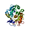
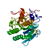
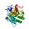
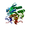
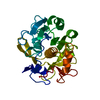
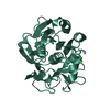
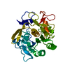
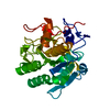
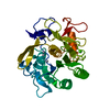
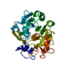
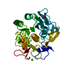
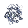
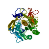
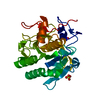
ProteinaseK / Endopeptidase K / Tritirachium alkaline proteinase
Mass: 28958.791 Da / Num. of mol.: 1 Source method: isolated from a genetically manipulated source Source: (gene. exp.) Parengyodontium album (fungus) / Gene: PROK / Production host: Parengyodontium album (fungus) / References: UniProt: P06873, peptidase K
In the structure databanks used in Yorodumi, some data are registered as the other names, "COVID-19 virus" and "2019-nCoV". Here are the details of the virus and the list of structure data.
Jan 31, 2019. EMDB accession codes are about to change! (news from PDBe EMDB page)
EMDB accession codes are about to change! (news from PDBe EMDB page)
The allocation of 4 digits for EMDB accession codes will soon come to an end. Whilst these codes will remain in use, new EMDB accession codes will include an additional digit and will expand incrementally as the available range of codes is exhausted. The current 4-digit format prefixed with “EMD-” (i.e. EMD-XXXX) will advance to a 5-digit format (i.e. EMD-XXXXX), and so on. It is currently estimated that the 4-digit codes will be depleted around Spring 2019, at which point the 5-digit format will come into force.
The EM Navigator/Yorodumi systems omit the EMD- prefix.
Related info.:Q: What is EMD? / ID/Accession-code notation in Yorodumi/EM Navigator
Yorodumi is a browser for structure data from EMDB, PDB, SASBDB, etc.
This page is also the successor to EM Navigator detail page, and also detail information page/front-end page for Omokage search.
The word "yorodu" (or yorozu) is an old Japanese word meaning "ten thousand". "mi" (miru) is to see.
Related info.:EMDB / PDB / SASBDB / Comparison of 3 databanks / Yorodumi Search / Aug 31, 2016. New EM Navigator & Yorodumi / Yorodumi Papers / Jmol/JSmol / Function and homology information / Changes in new EM Navigator and Yorodumi
 Movie
Movie Controller


 Open data
 Basic information
 Components
 Keywords
 Function and homology information
 Parengyodontium album (fungus)
 MOLECULAR REPLACEMENT /
  Authors
 United States,
  Brazil,
  Spain, 9items
  Citation
 Structure visualization
 Downloads & links
 Other downloads


 PDBj
PDBj

 Assembly
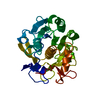

 Mass: 40.078 Da / Num. of mol.: 2 / Source method: obtained synthetically / Formula: Ca
 Mass: 18.015 Da / Num. of mol.: 121 / Source method: isolated from a natural source / Formula: H2O
 Sample preparation
 Processing