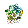
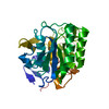
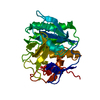
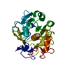
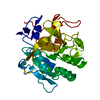
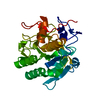
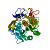
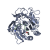
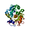
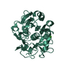
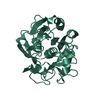
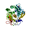
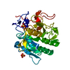
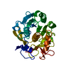
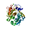
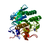
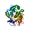
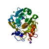
Journal: IUCrJ / Year: 2019 Title: MicroED with the Falcon III direct electron detector. Authors: Johan Hattne / Michael W Martynowycz / Pawel A Penczek / Tamir Gonen / Abstract: Microcrystal electron diffraction (MicroED) combines crystallography and electron cryo-microscopy (cryo-EM) into a method that is applicable to high-resolution structure determination. In MicroED, ...Microcrystal electron diffraction (MicroED) combines crystallography and electron cryo-microscopy (cryo-EM) into a method that is applicable to high-resolution structure determination. In MicroED, nanosized crystals, which are often intractable using other techniques, are probed by high-energy electrons in a transmission electron microscope. Diffraction data are recorded by a camera in movie mode: the nanocrystal is continuously rotated in the beam, thus creating a sequence of frames that constitute a movie with respect to the rotation angle. Until now, diffraction-optimized cameras have mostly been used for MicroED. Here, the use of a direct electron detector that was designed for imaging is reported. It is demonstrated that data can be collected more rapidly using the Falcon III for MicroED and with markedly lower exposure than has previously been reported. The Falcon III was operated at 40 frames per second and complete data sets reaching atomic resolution were recorded in minutes. The resulting density maps to 2.1 Å resolution of the serine protease proteinase K showed no visible signs of radiation damage. It is thus demonstrated that dedicated diffraction-optimized detectors are not required for MicroED, as shown by the fact that the very same cameras that are used for imaging applications in electron microscopy, such as single-particle cryo-EM, can also be used effectively for diffraction measurements.
Instrument: FEI VITROBOT MARK IV / Cryogen name: ETHANE / Humidity: 100 % / Chamber temperature: 298 K
-
Data collection
Experimental equipment
Model: Talos Arctica / Image courtesy: FEI Company
Microscopy
Model: FEI TALOS ARCTICA
Electron gun
Electron source: FIELD EMISSION GUN / Accelerating voltage: 200 kV / Illumination mode: FLOOD BEAM
Electron lens
Mode: DIFFRACTION
Specimen holder
Cryogen: NITROGEN / Specimen holder model: FEI TITAN KRIOS AUTOGRID HOLDER / Temperature (max): 100 K / Temperature (min): 77 K
Image recording
Average exposure time: 2.4069 sec. / Electron dose: 0.024069 e/Å2 / Film or detector model: FEI CETA (4k x 4k) / Num. of diffraction images: 165 / Num. of grids imaged: 1 / Num. of real images: 165
Image scans
Sampling size: 28 µm / Width: 2048 / Height: 2048
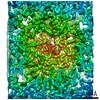
EM diffraction
Camera length: 2131 mm
EM diffraction shell
Resolution: 2.7→2.83 Å / Fourier space coverage: 97.2 % / Multiplicity: 3.8 / Num. of structure factors: 825 / Phase residual: 61.97 °
EM diffraction stats
Fourier space coverage: 98 % / High resolution: 2.7 Å / Num. of intensities measured: 28788 / Num. of structure factors: 6520 / Phase error: 43.59 ° / Phase residual: 43.59 ° / Phase error rejection criteria: 0 / Rmerge: 0.44 / Rsym: 0.44
Resolution: 2.7→2.7 Å / Cor.coef. Fo:Fc: 0.899 / Cor.coef. Fo:Fc free: 0.827 / SU B: 24.677 / SU ML: 0.443 / Cross valid method: THROUGHOUT / ESU R Free: 0.429 Details: Hydrogens have been added in their riding positions
Rfactor
Num. reflection
% reflection
Selection details
Rfree
0.2659
421
-
Copied from PDB entry 5K7S
Rwork
0.2313
-
-
-
all
0.234
-
-
-
obs
-
6483
97.621 %
-
Solvent computation
Ion probe radii: 0.8 Å / Shrinkage radii: 0.8 Å / VDW probe radii: 1.2 Å
 Movie
Movie Controller


 Open data
 Basic information
 Components
 Keywords
 Function and homology information
 Parengyodontium album (fungus)
 MOLECULAR REPLACEMENT / cryo EM / Resolution: 2.7 Å
  Authors
 Citation
 Structure visualization
 Downloads & links
 Other downloads
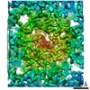


 PDBj
PDBj

 Assembly
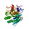

 Mass: 40.078 Da / Num. of mol.: 2 / Source method: obtained synthetically / Formula: Ca
 Mass: 18.015 Da / Num. of mol.: 3 / Source method: isolated from a natural source / Formula: H2O
 Sample preparation
 Processing