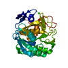
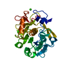
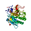
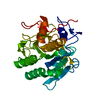
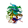
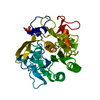
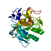
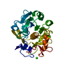
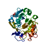
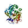
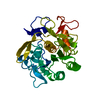
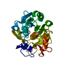
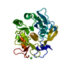
Entry Database : PDB / ID : 6mh6Title High-viscosity injector-based Pink Beam Serial Crystallography of Micro-crystals at a Synchrotron Radiation Source. Proteinase K Keywords / / Function / homology Function Domain/homology Component
/ / / / / / / / / / / / / / / / / / / / / / / / / / / / Biological species Parengyodontium album (fungus)Method / / / Resolution : 1.8 Å Authors Martin-Garcia, J.M. / Zhu, L. / Mendez, D. / Lee, M. / Chun, E. / Li, C. / Hu, H. / Subramanian, G. / Kissick, D. / Ogata, C. ...Martin-Garcia, J.M. / Zhu, L. / Mendez, D. / Lee, M. / Chun, E. / Li, C. / Hu, H. / Subramanian, G. / Kissick, D. / Ogata, C. / Henning, R. / Ishchenko, A. / Dobson, Z. / Zhan, S. / Weierstall, U. / Spence, J.C.H. / Fromme, P. / Zatsepin, N.A. / Fischetti, R.F. / Cherezov, V. / Liu, W. Funding support Organization Grant number Country National Science Foundation (NSF, United States) 1231306 National Science Foundation (NSF, United States) 1565180 National Institutes of Health/National Institute of General Medical Sciences (NIH/NIGMS) R21DA042298 National Institutes of Health/National Institute of General Medical Sciences (NIH/NIGMS) R01GM124152 National Institutes of Health/National Institute of General Medical Sciences (NIH/NIGMS) R01GM095583 National Institutes of Health/National Institute of General Medical Sciences (NIH/NIGMS) R35GM127086 National Institutes of Health/National Institute of General Medical Sciences (NIH/NIGMS) R24GM111072 National Institutes of Health/National Institute of General Medical Sciences (NIH/NIGMS) ACB-12002 National Institutes of Health/National Cancer Institute (NIH/NCI) AGM12006 Department of Energy (DOE, United States) DE-AC02-06CH11357
Journal : Iucrj / Year : 2019Title : High-viscosity injector-based pink-beam serial crystallography of microcrystals at a synchrotron radiation source.Authors: Martin-Garcia, J.M. / Zhu, L. / Mendez, D. / Lee, M.Y. / Chun, E. / Li, C. / Hu, H. / Subramanian, G. / Kissick, D. / Ogata, C. / Henning, R. / Ishchenko, A. / Dobson, Z. / Zhang, S. / ... Authors : Martin-Garcia, J.M. / Zhu, L. / Mendez, D. / Lee, M.Y. / Chun, E. / Li, C. / Hu, H. / Subramanian, G. / Kissick, D. / Ogata, C. / Henning, R. / Ishchenko, A. / Dobson, Z. / Zhang, S. / Weierstall, U. / Spence, J.C.H. / Fromme, P. / Zatsepin, N.A. / Fischetti, R.F. / Cherezov, V. / Liu, W. History Deposition Sep 17, 2018 Deposition site / Processing site Revision 1.0 Apr 24, 2019 Provider / Type Revision 1.1 May 29, 2019 Group / Database references / Category / citation_authorItem _citation.journal_volume / _citation.page_first ... _citation.journal_volume / _citation.page_first / _citation.page_last / _citation.pdbx_database_id_DOI / _citation.pdbx_database_id_PubMed / _citation.title / _citation_author.identifier_ORCID / _citation_author.name Revision 1.2 Aug 21, 2019 Group / Category / Item / _reflns.pdbx_Rmerge_I_obsRevision 1.3 Nov 6, 2019 Group / Data collection / Category Revision 1.4 Nov 27, 2019 Group / Category / Item Revision 1.5 Oct 11, 2023 Group Data collection / Database references ... Data collection / Database references / Derived calculations / Refinement description Category chem_comp_atom / chem_comp_bond ... chem_comp_atom / chem_comp_bond / database_2 / pdbx_initial_refinement_model / pdbx_struct_conn_angle / struct_conn Item _database_2.pdbx_DOI / _database_2.pdbx_database_accession ... _database_2.pdbx_DOI / _database_2.pdbx_database_accession / _pdbx_struct_conn_angle.ptnr1_auth_seq_id / _pdbx_struct_conn_angle.ptnr3_auth_seq_id / _pdbx_struct_conn_angle.value / _struct_conn.pdbx_dist_value / _struct_conn.ptnr2_auth_seq_id Revision 1.6 Oct 16, 2024 Group / Category / pdbx_modification_feature
Show all Show less
 Movie
Movie Controller

 Yorodumi
 Open data
 Basic information
 Components
 Keywords
 Function and homology information
 Parengyodontium album (fungus)
 X-RAY DIFFRACTION /
  Authors
 United States, 10items
  Citation
 Structure visualization
 Downloads & links
 Other downloads


 PDBj
PDBj

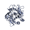
 Assembly
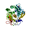

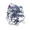
 Mass: 40.078 Da / Num. of mol.: 2 / Source method: obtained synthetically / Formula: Ca
 Mass: 62.005 Da / Num. of mol.: 1 / Source method: obtained synthetically / Formula: NO3
 Mass: 18.015 Da / Num. of mol.: 137 / Source method: isolated from a natural source / Formula: H2O
 Sample preparation
 Processing