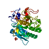
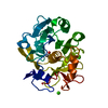
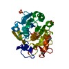
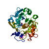
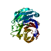
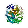
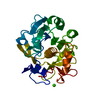
[English] 日本語
Yorodumi
- PDB-5wjg: Using sound pulses to solve the crystal harvesting bottleneck -

+
Open data


ID or keywords:

Loading...

-
Basic information

Entry
Database: PDB / ID: 5wjg
TitleUsing sound pulses to solve the crystal harvesting bottleneck
ComponentsProteinase K
KeywordsHYDROLASE / Crystal harvesting / drug discovery / acoustic droplet ejection
Function / homology
Function and homology information


peptidase K / serine-type endopeptidase activity / proteolysis / extracellular region / metal ion binding
Similarity search - Function
Proteinase K-like catalytic domain / Peptidase S8/S53 domain / Peptidase S8 propeptide/proteinase inhibitor I9 / Peptidase inhibitor I9 / Peptidase S8 propeptide/proteinase inhibitor I9 superfamily / Peptidase S8, subtilisin, His-active site / Serine proteases, subtilase family, histidine active site. / Serine proteases, subtilase family, aspartic acid active site. / Peptidase S8, subtilisin, Asp-active site / Serine proteases, subtilase family, serine active site. ...Proteinase K-like catalytic domain / Peptidase S8/S53 domain / Peptidase S8 propeptide/proteinase inhibitor I9 / Peptidase inhibitor I9 / Peptidase S8 propeptide/proteinase inhibitor I9 superfamily / Peptidase S8, subtilisin, His-active site / Serine proteases, subtilase family, histidine active site. / Serine proteases, subtilase family, aspartic acid active site. / Peptidase S8, subtilisin, Asp-active site / Serine proteases, subtilase family, serine active site. / Peptidase S8, subtilisin, Ser-active site / Serine proteases, subtilase domain profile. / Peptidase S8, subtilisin-related / Peptidase S8/S53 domain superfamily / Subtilase family / Peptidase S8/S53 domain / Rossmann fold / 3-Layer(aba) Sandwich / Alpha Beta
Similarity search - Domain/homology
L(+)-TARTARIC ACID / Proteinase K
Similarity search - Component
Biological speciesParengyodontium album (fungus)
MethodX-RAY DIFFRACTION / SYNCHROTRON / MOLECULAR REPLACEMENT / Resolution: 1.5 Å
AuthorsSoares, A.S. / Brennan, H.M. / McCarthy, L. / Leroy, L.
Funding support United States, 4items
OrganizationGrant numberCountry
National Institutes of Health/National Institute of General Medical Sciences (NIH/NIGMS)P41GM111244 United States
Department of Energy (DOE, United States)KP1605010 United States
Office of Workforce Development for Teachers and Scientists (WDTS) United States
National Science Foundation (NSF, United States)P41GM103393 United States
CitationJournal: Acta Crystallogr D Struct Biol / Year: 2018
Title: Using sound pulses to solve the crystal-harvesting bottleneck.
Authors: Samara, Y.N. / Brennan, H.M. / McCarthy, L. / Bollard, M.T. / Laspina, D. / Wlodek, J.M. / Campos, S.L. / Natarajan, R. / Gofron, K. / McSweeney, S. / Soares, A.S. / Leroy, L.
History
DepositionJul 22, 2017Deposition site: RCSB / Processing site: RCSB
Revision 1.0Aug 2, 2017Provider: repository / Type: Initial release
Revision 1.1Sep 20, 2017Group: Author supporting evidence / Category: pdbx_audit_support / Item: _pdbx_audit_support.funding_organization
Revision 1.2Oct 17, 2018Group: Data collection / Database references / Category: citation / citation_author
Item: _citation.journal_abbrev / _citation.journal_id_CSD ..._citation.journal_abbrev / _citation.journal_id_CSD / _citation.journal_id_ISSN / _citation.journal_volume / _citation.page_first / _citation.page_last / _citation.pdbx_database_id_DOI / _citation.pdbx_database_id_PubMed / _citation.title / _citation.year
Revision 1.3Nov 27, 2019Group: Author supporting evidence / Category: pdbx_audit_support / Item: _pdbx_audit_support.funding_organization
Revision 1.4Oct 4, 2023Group: Advisory / Data collection ...Advisory / Data collection / Database references / Refinement description
Category: chem_comp_atom / chem_comp_bond ...chem_comp_atom / chem_comp_bond / database_2 / pdbx_initial_refinement_model / pdbx_unobs_or_zero_occ_atoms
Item: _database_2.pdbx_DOI / _database_2.pdbx_database_accession

-
Structure visualization

Structure viewerMolecule:
MolmilJmol/JSmol

Downloads & links

-
Assembly

Deposited unit
A: Proteinase K
hetero molecules


Theoretical massNumber of molelcules
Total (without water)29,2735
Polymers28,9591
Non-polymers3144
Water8,647480
1


  • Idetical with deposited unit
  • defined by author&software
TypeNameSymmetry operationNumber
identity operation1_555x,y,z1
MethodPISA
Unit cell
Length a, b, c (Å)68.041, 68.041, 102.286
Angle α, β, γ (deg.)90.00, 90.00, 90.00
Int Tables number96
Space group name H-MP43212
Components on special symmetry positions
IDModelComponents
11A-546-

HOH

21A-743-

HOH

-
Components

#1: Protein Proteinase K / / Endopeptidase K / Tritirachium alkaline proteinase


Mass: 28958.791 Da / Num. of mol.: 1 / Source method: isolated from a natural source / Source: (natural) Parengyodontium album (fungus) / References: UniProt: P06873, peptidase K
#2: Chemical ChemComp-CA / CALCIUM ION


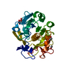
Mass: 40.078 Da / Num. of mol.: 1 / Source method: obtained synthetically / Formula: Ca
#3: Chemical ChemComp-TLA / L(+)-TARTARIC ACID / Tartaric acid


Mass: 150.087 Da / Num. of mol.: 1 / Source method: obtained synthetically / Formula: C4H6O6 / Feature type: SUBJECT OF INVESTIGATION
#4: Chemical ChemComp-EDO / 1,2-ETHANEDIOL / ETHYLENE GLYCOL / Ethylene glycol


Mass: 62.068 Da / Num. of mol.: 2 / Source method: obtained synthetically / Formula: C2H6O2
#5: Water ChemComp-HOH / water / Water


Mass: 18.015 Da / Num. of mol.: 480 / Source method: isolated from a natural source / Formula: H2O

-
Experimental details

-
Experiment

ExperimentMethod: X-RAY DIFFRACTION / Number of used crystals: 1

-
Sample preparation

CrystalDensity Matthews: 2.04 Å3/Da / Density % sol: 39.82 %
Crystal growTemperature: 295 K / Method: batch mode / pH: 6.5 / Details: 0.1 M BisTris pH 6.5, 0.08M CaCl2

-
Data collection

DiffractionMean temperature: 100 K
Diffraction sourceSource: SYNCHROTRON / Site: NSLS-II / Beamline: 17-ID-1 / Wavelength: 0.97913 Å
DetectorType: DECTRIS EIGER X 9M / Detector: PIXEL / Date: Jun 16, 2017
RadiationProtocol: SINGLE WAVELENGTH / Monochromatic (M) / Laue (L): M / Scattering type: x-ray
Radiation wavelengthWavelength: 0.97913 Å / Relative weight: 1
ReflectionResolution: 1.5→102.29 Å / Num. obs: 36472 / % possible obs: 97.88 % / Redundancy: 10.2 % / Rmerge(I) obs: 0.076 / Net I/σ(I): 21.8

-
Processing

Software
NameVersionClassification
REFMAC5.8.0073refinement
XDSdata reduction
XDSdata scaling
MOLREPphasing
RefinementMethod to determine structure: MOLECULAR REPLACEMENT
Starting model: 4fon
Resolution: 1.5→102.29 Å / Cor.coef. Fo:Fc: 0.974 / Cor.coef. Fo:Fc free: 0.964 / SU B: 0.938 / SU ML: 0.035 / Cross valid method: THROUGHOUT / ESU R: 0.06 / ESU R Free: 0.065 / Details: HYDROGENS HAVE BEEN ADDED IN THE RIDING POSITIONS
RfactorNum. reflection% reflectionSelection details
Rfree0.15736 1896 4.9 %RANDOM
Rwork0.12366 ---
obs0.12528 36472 97.88 %-
Solvent computationIon probe radii: 0.8 Å / Shrinkage radii: 0.8 Å / VDW probe radii: 1.2 Å
Displacement parametersBiso mean: 8.694 Å2
Baniso -1Baniso -2Baniso -3
1--0 Å20 Å20 Å2
2---0 Å20 Å2
3---0 Å2
Refinement stepCycle: 1 / Resolution: 1.5→102.29 Å
ProteinNucleic acidLigandSolventTotal
Num. atoms2032 0 19 480 2531
Refine LS restraints
Refine-IDTypeDev idealDev ideal targetNumber
X-RAY DIFFRACTIONr_bond_refined_d0.0250.0192168
X-RAY DIFFRACTIONr_bond_other_d0.0070.021944
X-RAY DIFFRACTIONr_angle_refined_deg2.2081.9422961
X-RAY DIFFRACTIONr_angle_other_deg1.03834481
X-RAY DIFFRACTIONr_dihedral_angle_1_deg6.3435302
X-RAY DIFFRACTIONr_dihedral_angle_2_deg37.00423.70889
X-RAY DIFFRACTIONr_dihedral_angle_3_deg11.51915317
X-RAY DIFFRACTIONr_dihedral_angle_4_deg18.0971513
X-RAY DIFFRACTIONr_chiral_restr0.1480.2327
X-RAY DIFFRACTIONr_gen_planes_refined0.0130.022599
X-RAY DIFFRACTIONr_gen_planes_other0.0020.02518
X-RAY DIFFRACTIONr_nbd_refined
X-RAY DIFFRACTIONr_nbd_other
X-RAY DIFFRACTIONr_nbtor_refined
X-RAY DIFFRACTIONr_nbtor_other
X-RAY DIFFRACTIONr_xyhbond_nbd_refined
X-RAY DIFFRACTIONr_xyhbond_nbd_other
X-RAY DIFFRACTIONr_metal_ion_refined
X-RAY DIFFRACTIONr_metal_ion_other
X-RAY DIFFRACTIONr_symmetry_vdw_refined
X-RAY DIFFRACTIONr_symmetry_vdw_other
X-RAY DIFFRACTIONr_symmetry_hbond_refined
X-RAY DIFFRACTIONr_symmetry_hbond_other
X-RAY DIFFRACTIONr_symmetry_metal_ion_refined
X-RAY DIFFRACTIONr_symmetry_metal_ion_other
X-RAY DIFFRACTIONr_mcbond_it0.8090.5641148
X-RAY DIFFRACTIONr_mcbond_other0.7910.5611147
X-RAY DIFFRACTIONr_mcangle_it1.1750.8461443
X-RAY DIFFRACTIONr_mcangle_other1.1770.8481444
X-RAY DIFFRACTIONr_scbond_it2.0810.7671020
X-RAY DIFFRACTIONr_scbond_other2.060.7671020
X-RAY DIFFRACTIONr_scangle_it
X-RAY DIFFRACTIONr_scangle_other3.0061.0791510
X-RAY DIFFRACTIONr_long_range_B_refined6.5397.7832989
X-RAY DIFFRACTIONr_long_range_B_other5.4955.9462683
X-RAY DIFFRACTIONr_rigid_bond_restr
X-RAY DIFFRACTIONr_sphericity_free
X-RAY DIFFRACTIONr_sphericity_bonded
LS refinement shellResolution: 1.5→1.539 Å / Total num. of bins used: 20
RfactorNum. reflection% reflection
Rfree0.29 112 -
Rwork0.213 2262 -
obs--83.21 %

+
About Yorodumi

-
News

-
Feb 9, 2022. New format data for meta-information of EMDB entries

New format data for meta-information of EMDB entries

  • Version 3 of the EMDB header file is now the official format.
  • The previous official version 1.9 will be removed from the archive.

Related info.:EMDB header

External links:wwPDB to switch to version 3 of the EMDB data model

-
Aug 12, 2020. Covid-19 info

Covid-19 info

URL: https://pdbj.org/emnavi/covid19.php

New page: Covid-19 featured information page in EM Navigator.

Related info.:Covid-19 info / Mar 5, 2020. Novel coronavirus structure data

+
Mar 5, 2020. Novel coronavirus structure data

Novel coronavirus structure data

Related info.:Yorodumi Speices / Aug 12, 2020. Covid-19 info

External links:COVID-19 featured content - PDBj / Molecule of the Month (242):Coronavirus Proteases

+
Jan 31, 2019. EMDB accession codes are about to change! (news from PDBe EMDB page)

EMDB accession codes are about to change! (news from PDBe EMDB page)

  • The allocation of 4 digits for EMDB accession codes will soon come to an end. Whilst these codes will remain in use, new EMDB accession codes will include an additional digit and will expand incrementally as the available range of codes is exhausted. The current 4-digit format prefixed with “EMD-” (i.e. EMD-XXXX) will advance to a 5-digit format (i.e. EMD-XXXXX), and so on. It is currently estimated that the 4-digit codes will be depleted around Spring 2019, at which point the 5-digit format will come into force.
  • The EM Navigator/Yorodumi systems omit the EMD- prefix.

Related info.:Q: What is EMD? / ID/Accession-code notation in Yorodumi/EM Navigator

External links:EMDB Accession Codes are Changing Soon! / Contact to PDBj

+
Jul 12, 2017. Major update of PDB

Major update of PDB

  • wwPDB released updated PDB data conforming to the new PDBx/mmCIF dictionary.
  • This is a major update changing the version number from 4 to 5, and with Remediation, in which all the entries are updated.
  • In this update, many items about electron microscopy experimental information are reorganized (e.g. em_software).
  • Now, EM Navigator and Yorodumi are based on the updated data.

External links:wwPDB Remediation / Enriched Model Files Conforming to OneDep Data Standards Now Available in the PDB FTP Archive

-
Yorodumi

Thousand views of thousand structures

  • Yorodumi is a browser for structure data from EMDB, PDB, SASBDB, etc.
  • This page is also the successor to EM Navigator detail page, and also detail information page/front-end page for Omokage search.
  • The word "yorodu" (or yorozu) is an old Japanese word meaning "ten thousand". "mi" (miru) is to see.

Related info.:EMDB / PDB / SASBDB / Comparison of 3 databanks / Yorodumi Search / Aug 31, 2016. New EM Navigator & Yorodumi / Yorodumi Papers / Jmol/JSmol / Function and homology information / Changes in new EM Navigator and Yorodumi

Read more