 Movie
Movie Controller

[English] 日本語
 Yorodumi
- PDB-3f7o: Crystal structure of Cuticle-Degrading Protease from Paecilomyces... -
+ Open data

- Basic information
| Entry | Database: PDB / ID: 3f7o | |||||||||
|---|---|---|---|---|---|---|---|---|---|---|
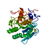
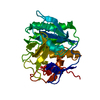
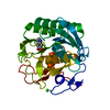
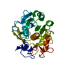
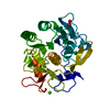
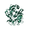
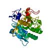
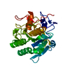
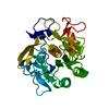
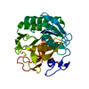
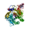
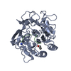
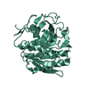
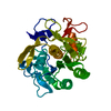
| Title | Crystal structure of Cuticle-Degrading Protease from Paecilomyces lilacinus (PL646) | |||||||||
 Components Components |
| |||||||||
 Keywords Keywords | HYDROLASE / Cuticle-Degrading Protease / Paecilomyces lilacinus / Protease / Serine protease | |||||||||
| Function / homology |  Function and homology information Function and homology informationserine-type endopeptidase activity / proteolysis / extracellular region / metal ion binding Similarity search - Function | |||||||||
| Biological species |  Purpureocillium lilacinum (fungus) Purpureocillium lilacinum (fungus)synthetic construct (others) | |||||||||
| Method |  X-RAY DIFFRACTION / MOLECULAR REPLACEMENT / Resolution: 2.2 Å X-RAY DIFFRACTION / MOLECULAR REPLACEMENT / Resolution: 2.2 Å | |||||||||
 Authors Authors | Liang, L. / Lou, Z. / Meng, Z. / Rao, Z. / Zhang, K. | |||||||||
 Citation Citation | Journal: Faseb J. / Year: 2010 Title: The crystal structures of two cuticle-degrading proteases from nematophagous fungi and their contribution to infection against nematodes. Authors: Liang, L. / Meng, Z. / Ye, F. / Yang, J. / Liu, S. / Sun, Y. / Guo, Y. / Mi, Q. / Huang, X. / Zou, C. / Rao, Z. / Lou, Z. / Zhang, K.Q. | |||||||||
| History |
|
- Structure visualization
| Structure viewer | Molecule: MolmilJmol/JSmol |
|---|
- Downloads & links
-Download
| PDBx/mmCIF format | 3f7o.cif.gz | 125.2 KB | Display | PDBx/mmCIF format |
|---|---|---|---|---|
| PDB format | pdb3f7o.ent.gz | 95.8 KB | Display | PDB format |
| PDBx/mmJSON format | 3f7o.json.gz | Tree view | PDBx/mmJSON format | |
| Others |  Other downloads Other downloads |
-Validation report
| Arichive directory | https://data.pdbj.org/pub/pdb/validation_reports/f7/3f7oftp://data.pdbj.org/pub/pdb/validation_reports/f7/3f7o | HTTPS FTP |
|---|
-Related structure data
| Related structure data | 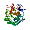 3f7mC 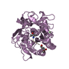 3prkS C: citing same article ( S: Starting model for refinement |
|---|---|
| Similar structure data |


-Links
 PDBj
PDBj

- Assembly
| Deposited unit | 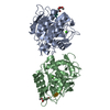
| ||||||||
|---|---|---|---|---|---|---|---|---|---|
| 1 | 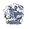
| ||||||||
| 2 | 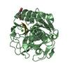
| ||||||||
| Unit cell |
|
-Components
| #1: Protein | Mass: 28743.723 Da / Num. of mol.: 2 / Source method: isolated from a natural source / Source: (natural) Purpureocillium lilacinum (fungus) / Plasmid: pMD18-T / References: UniProt: Q01471#2: Protein/peptide | Mass: 470.516 Da / Num. of mol.: 2 / Source method: obtained synthetically / Source: (synth.) synthetic construct (others) #3: Chemical |   Mass: 40.078 Da / Num. of mol.: 2 / Source method: obtained synthetically / Formula: Ca Mass: 40.078 Da / Num. of mol.: 2 / Source method: obtained synthetically / Formula: Ca#4: Water | ChemComp-HOH / |  Mass: 18.015 Da / Num. of mol.: 517 / Source method: isolated from a natural source / Formula: H2O Mass: 18.015 Da / Num. of mol.: 517 / Source method: isolated from a natural source / Formula: H2OHas protein modification | Y | |
|---|
-Experimental details
-Experiment
| Experiment | Method: X-RAY DIFFRACTION / Number of used crystals: 1 |
|---|
- Sample preparation
| Crystal | Density Matthews: 2.36 Å3/Da / Density % sol: 47.89 % |
|---|---|
| Crystal grow | Temperature: 289 K / Method: vapor diffusion, hanging drop / pH: 6 Details: 20% PEG 4000, pH 6.0, VAPOR DIFFUSION, HANGING DROP, temperature 289K |
-Data collection
| Diffraction | Mean temperature: 100 K |
|---|---|
| Diffraction source | Source: ROTATING ANODE / Type: RIGAKU MICROMAX-007 / Wavelength: 1.5418 Å |
| Detector | Type: RIGAKU RAXIS IV++ / Detector: IMAGE PLATE / Date: Jul 5, 2007 / Details: osmic mirror |
| Radiation | Monochromator: osmic mirror / Protocol: SINGLE WAVELENGTH / Monochromatic (M) / Laue (L): M / Scattering type: x-ray |
| Radiation wavelength | Wavelength: 1.5418 Å / Relative weight: 1 |
| Reflection | Resolution: 2.1→50 Å / Num. obs: 31500 / % possible obs: 95.7 % / Observed criterion σ(F): 0 / Redundancy: 3.9 % / Rmerge(I) obs: 0.136 |
| Reflection shell | Resolution: 2.1→2.18 Å / Redundancy: 2.8 % / Rmerge(I) obs: 0.564 / Num. unique all: 2806 / % possible all: 85.7 |
- Processing
| Software |
| |||||||||||||||||||||||||
|---|---|---|---|---|---|---|---|---|---|---|---|---|---|---|---|---|---|---|---|---|---|---|---|---|---|---|
| Refinement | Method to determine structure: MOLECULAR REPLACEMENT Starting model: 3PRK Resolution: 2.2→50 Å / σ(F): 0 / Stereochemistry target values: Engh & Huber
| |||||||||||||||||||||||||
| Refinement step | Cycle: LAST / Resolution: 2.2→50 Å
| |||||||||||||||||||||||||
| Refine LS restraints |
|
