 Movie
Movie Controller

[English] 日本語
 Yorodumi
- PDB-3prk: INHIBITION OF PROTEINASE K BY METHOXYSUCCINYL-ALA-ALA-PRO-ALA-CHL... -
+ Open data

- Basic information
| Entry | Database: PDB / ID: 3prk | ||||||
|---|---|---|---|---|---|---|---|
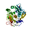
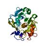
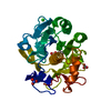
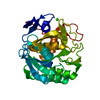
| Title | INHIBITION OF PROTEINASE K BY METHOXYSUCCINYL-ALA-ALA-PRO-ALA-CHLOROMETHYL KETONE. AN X-RAY STUDY AT 2.2-ANGSTROMS RESOLUTION | ||||||
 Components Components |
| ||||||
 Keywords Keywords | HYDROLASE/HYDROLASE INHIBITOR / SERINE PROTEINASE / HYDROLASE-HYDROLASE INHIBITOR COMPLEX | ||||||
| Function / homology |  Function and homology information Function and homology informationpeptidase K / serine-type endopeptidase activity / proteolysis / extracellular region / metal ion binding Similarity search - Function | ||||||
| Biological species |  Engyodontium album (fungus) Engyodontium album (fungus) | ||||||
| Method |  X-RAY DIFFRACTION / Resolution: 2.2 Å X-RAY DIFFRACTION / Resolution: 2.2 Å | ||||||
 Authors Authors | Wolf, W.M. / Bajorath, J. / Mueller, A. / Raghunathan, S. / Singh, T.P. / Hinrichs, W. / Saenger, W. | ||||||
 Citation Citation | Journal: J.Biol.Chem. / Year: 1991 Title: Inhibition of proteinase K by methoxysuccinyl-Ala-Ala-Pro-Ala-chloromethyl ketone. An x-ray study at 2.2-A resolution. Authors: Wolf, W.M. / Bajorath, J. / Muller, A. / Raghunathan, S. / Singh, T.P. / Hinrichs, W. / Saenger, W. #1: Journal: Acta Crystallogr.,Sect.B / Year: 1988Title: Synchrotron X-Ray Data Collection and Restrained Least-Squares Refinement of the Crystal Structure of Proteinase K at 1.5 Angstroms Resolution Authors: Betzel, C. / Pal, G.P. / Saenger, W. #2: Journal: FEBS Lett. / Year: 1986Title: Crystallization of the Bifunctional Proteinase(Slash)Amylase Inhibitor Pki-3 and of its Complex with Proteinase K Authors: Pal, G.P. / Betzel, C. / Jany, K.-D. / Saenger, W. #3: Journal: FEBS Lett. / Year: 1986Title: Active-Site Geometry of Proteinase K. Crystallographic Study of its Complex with a Dipeptide Chloromethyl Ketone Inhibitor Authors: Betzel, C. / Pal, G.P. / Struck, M. / Jany, K.-D. / Saenger, W. #4: Journal: Embo J. / Year: 1984Title: Three-Dimensional Structure of Fungal Proteinase K Reveals Similarity to Bacterial Subtilisin Authors: Paehler, A. / Banerjee, A. / Dattagupta, J.K. / Fujiwara, T. / Lindner, K. / Pal, G.P. / Suck, D. / Weber, G. / Saenger, W. #5: Journal: J.Mol.Biol. / Year: 1975Title: Crystallization of the Fungal Enzyme Proteinase K and Amino Acid Composition Authors: Dattagupta, J.K. / Fujiwara, T. / Grishin, E.V. / Lindner, K. / Manor, P.C. / Pieniazek, N.J. / Saenger, W. / Suck, D. | ||||||
| History |
|
- Structure visualization
| Structure viewer | Molecule: MolmilJmol/JSmol |
|---|
- Downloads & links
-Download
| PDBx/mmCIF format | 3prk.cif.gz | 69.2 KB | Display | PDBx/mmCIF format |
|---|---|---|---|---|
| PDB format | pdb3prk.ent.gz | 50.1 KB | Display | PDB format |
| PDBx/mmJSON format | 3prk.json.gz | Tree view | PDBx/mmJSON format | |
| Others |  Other downloads Other downloads |
-Validation report
| Arichive directory | https://data.pdbj.org/pub/pdb/validation_reports/pr/3prkftp://data.pdbj.org/pub/pdb/validation_reports/pr/3prk | HTTPS FTP |
|---|
-Related structure data
| Similar structure data |
|---|


-Links
 PDBj
PDBj

- Assembly
| Deposited unit | 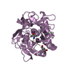
| |||||||||
|---|---|---|---|---|---|---|---|---|---|---|
| 1 |
| |||||||||
| Unit cell |
| |||||||||
| Atom site foot note | 1: CIS PROLINE - PRO E 171 | |||||||||
| Components on special symmetry positions |
|
-Components
| #1: Protein | Mass: 28930.783 Da / Num. of mol.: 1 Source method: isolated from a genetically manipulated source Source: (gene. exp.) Engyodontium album (fungus) / Tissue: limber / Gene: PROK / References: UniProt: P06873, peptidase K |
|---|---|
| #2: Protein/peptide | 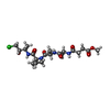  Type: Peptide-like / Class: Inhibitor / Mass: 476.952 Da / Num. of mol.: 1 / Source method: obtained synthetically Type: Peptide-like / Class: Inhibitor / Mass: 476.952 Da / Num. of mol.: 1 / Source method: obtained syntheticallyReferences: methoxysuccinyl-alanyl-alanyl-prolyl-alanine chloromethyl ketone |
| #3: Chemical | ChemComp-CA /   Mass: 40.078 Da / Num. of mol.: 1 / Source method: obtained synthetically / Formula: Ca Mass: 40.078 Da / Num. of mol.: 1 / Source method: obtained synthetically / Formula: Ca |
| #4: Water | ChemComp-HOH /  Mass: 18.015 Da / Num. of mol.: 170 / Source method: isolated from a natural source / Formula: H2O Mass: 18.015 Da / Num. of mol.: 170 / Source method: isolated from a natural source / Formula: H2O |
| Compound details | THE CHLOROMETHYLKETONE GROUP IS COVALENTLY LINKED WITH THE ACTIVE SITE FUNCTIONAL GROUPS NE2 HIS E ...THE CHLOROMETH |
| Has protein modification | Y |
-Experimental details
-Experiment
| Experiment | Method: X-RAY DIFFRACTION |
|---|
- Sample preparation
| Crystal | Density Matthews: 2.15 Å3/Da / Density % sol: 42.83 % | ||||||||||||||||||||||||||||||||||||
|---|---|---|---|---|---|---|---|---|---|---|---|---|---|---|---|---|---|---|---|---|---|---|---|---|---|---|---|---|---|---|---|---|---|---|---|---|---|
| Crystal grow | *PLUS pH: 7.5 / Method: vapor diffusion, sitting drop | ||||||||||||||||||||||||||||||||||||
| Components of the solutions | *PLUS
|
-Data collection
| Radiation | Scattering type: x-ray |
|---|---|
| Radiation wavelength | Relative weight: 1 |
| Reflection | *PLUS Highest resolution: 2.2 Å / Num. obs: 11864 / Observed criterion σ(F): 1 / Num. measured all: 53620 / Rmerge F obs: 0.098 / Biso Wilson estimate: 14.6 Å2 |
- Processing
| Software | Name: TNT / Classification: refinement | ||||||||||||||||||||||||||||||
|---|---|---|---|---|---|---|---|---|---|---|---|---|---|---|---|---|---|---|---|---|---|---|---|---|---|---|---|---|---|---|---|
| Refinement | Rfactor obs: 0.198 / Highest resolution: 2.2 Å | ||||||||||||||||||||||||||||||
| Refinement step | Cycle: LAST / Highest resolution: 2.2 Å
| ||||||||||||||||||||||||||||||
| Refine LS restraints |
| ||||||||||||||||||||||||||||||
| Software | *PLUS Name: TNT / Classification: refinement | ||||||||||||||||||||||||||||||
| Refinement | *PLUS Highest resolution: 2.2 Å / Lowest resolution: 10 Å / Num. reflection obs: 11864 / Rfactor obs: 0.198 | ||||||||||||||||||||||||||||||
| Solvent computation | *PLUS | ||||||||||||||||||||||||||||||
| Displacement parameters | *PLUS Biso mean: 15 Å2 | ||||||||||||||||||||||||||||||
| Refine LS restraints | *PLUS
|
