 Movie
Movie Controller

[English] 日本語
 Yorodumi
- PDB-1bjr: COMPLEX FORMED BETWEEN PROTEOLYTICALLY GENERATED LACTOFERRIN FRAG... -
+ Open data

- Basic information
| Entry | Database: PDB / ID: 1bjr | ||||||
|---|---|---|---|---|---|---|---|
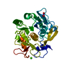
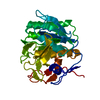
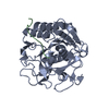
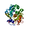
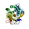
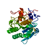
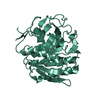
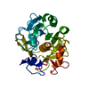
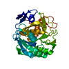
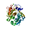
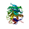
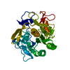
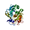
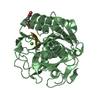
| Title | COMPLEX FORMED BETWEEN PROTEOLYTICALLY GENERATED LACTOFERRIN FRAGMENT AND PROTEINASE K | ||||||
 Components Components |
| ||||||
 Keywords Keywords | HYDROLASE/HYDROLASE INHIBITOR / PROTEINASE K / LACTOFERRIN / IRON TRANSPORT / HYDROLASE-HYDROLASE INHIBITOR complex | ||||||
| Function / homology |  Function and homology information Function and homology informationpeptidase K / serine-type endopeptidase activity / proteolysis / extracellular region / metal ion binding Similarity search - Function | ||||||
| Biological species |  Engyodontium album (fungus) Engyodontium album (fungus) Bubalus bubalis (water buffalo) Bubalus bubalis (water buffalo) | ||||||
| Method |  X-RAY DIFFRACTION / MOLECULAR REPLACEMENT / Resolution: 2.44 Å X-RAY DIFFRACTION / MOLECULAR REPLACEMENT / Resolution: 2.44 Å | ||||||
 Authors Authors | Singh, T.P. / Sharma, S. / Karthikeyan, S. / Betzel, C. / Bhatia, K.L. | ||||||
 Citation Citation | Journal: Proteins / Year: 1998 Title: Crystal structure of a complex formed between proteolytically-generated lactoferrin fragment and proteinase K. Authors: Singh, T.P. / Sharma, S. / Karthikeyan, S. / Betzel, C. / Bhatia, K.L. #1: Journal: J.Biol.Chem. / Year: 1991Title: Inhibition of Proteinase K by Methoxysuccinyl-Ala-Ala-Pro-Ala-Chloromethyl Ketone. An X-Ray Study at 2.2-A Resolution Authors: Wolf, W.M. / Bajorath, J. / Muller, A. / Raghunathan, S. / Singh, T.P. / Hinrichs, W. / Saenger, W. #2: Journal: Acta Crystallogr.,Sect.B / Year: 1988Title: Synchrotron X-Ray Data Collection and Restrained Least-Squares Refinement of the Crystal Structure of Proteinase K at 1.5 A Resolution Authors: Betzel, C. / Pal, G.P. / Saenger, W. | ||||||
| History |
|
- Structure visualization
| Structure viewer | Molecule: MolmilJmol/JSmol |
|---|
- Downloads & links
-Download
| PDBx/mmCIF format | 1bjr.cif.gz | 71.9 KB | Display | PDBx/mmCIF format |
|---|---|---|---|---|
| PDB format | pdb1bjr.ent.gz | 51.8 KB | Display | PDB format |
| PDBx/mmJSON format | 1bjr.json.gz | Tree view | PDBx/mmJSON format | |
| Others |  Other downloads Other downloads |
-Validation report
| Arichive directory | https://data.pdbj.org/pub/pdb/validation_reports/bj/1bjrftp://data.pdbj.org/pub/pdb/validation_reports/bj/1bjr | HTTPS FTP |
|---|
-Related structure data
| Related structure data |  3prkS S: Starting model for refinement |
|---|---|
| Similar structure data |

-Links
 PDBj
PDBj

- Assembly
| Deposited unit | 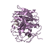
| ||||||||
|---|---|---|---|---|---|---|---|---|---|
| 1 |
| ||||||||
| Unit cell |
|
-Components
| #1: Protein | Mass: 28930.783 Da / Num. of mol.: 1 / Source method: isolated from a natural source / Source: (natural) Engyodontium album (fungus) / Strain: LIMBER / References: UniProt: P06873, peptidase K | ||||
|---|---|---|---|---|---|
| #2: Protein/peptide | Mass: 813.899 Da / Num. of mol.: 1 / Fragment: FRAGMENT / Source method: isolated from a natural source / Source: (natural) Bubalus bubalis (water buffalo) | ||||
| #3: Chemical |   Mass: 40.078 Da / Num. of mol.: 2 / Source method: obtained synthetically / Formula: Ca Mass: 40.078 Da / Num. of mol.: 2 / Source method: obtained synthetically / Formula: Ca#4: Water | ChemComp-HOH / |  Mass: 18.015 Da / Num. of mol.: 201 / Source method: isolated from a natural source / Formula: H2O Mass: 18.015 Da / Num. of mol.: 201 / Source method: isolated from a natural source / Formula: H2OHas protein modification | Y | |
-Experimental details
-Experiment
| Experiment | Method: X-RAY DIFFRACTION / Number of used crystals: 1 |
|---|
- Sample preparation
| Crystal | Density Matthews: 2.19 Å3/Da / Density % sol: 43.7 % | |||||||||||||||
|---|---|---|---|---|---|---|---|---|---|---|---|---|---|---|---|---|
| Crystal grow | Temperature: 279 K / Method: microdialysis / pH: 6 Details: 60 MG/ML PROTEIN IN 10MM TRIS.HCL, PH 6.0 WAS MICRODIALYZED AGAINST 10% ETHANOL AT 6 DEGREE CELSIUS, microdialysis, temperature 279K | |||||||||||||||
| Crystal | *PLUS | |||||||||||||||
| Crystal grow | *PLUS Method: microdialysis | |||||||||||||||
| Components of the solutions | *PLUS
|
-Data collection
| Diffraction | Mean temperature: 288 K |
|---|---|
| Diffraction source | Source: ROTATING ANODE / Type: RIGAKU RUH2R / Wavelength: 1.5418 |
| Detector | Type: MARRESEARCH / Detector: IMAGE PLATE / Date: Feb 1, 1997 / Details: PIN HOLE |
| Radiation | Monochromator: GRAPHITE(002) / Monochromatic (M) / Laue (L): M / Scattering type: x-ray |
| Radiation wavelength | Wavelength: 1.5418 Å / Relative weight: 1 |
| Reflection | Resolution: 2.44→12 Å / Num. obs: 9051 / % possible obs: 92.2 % / Observed criterion σ(I): 3 / Redundancy: 2.42 % / Biso Wilson estimate: 29.19 Å2 / Rmerge(I) obs: 0.07 / Rsym value: 0.07 / Net I/σ(I): 22.2 |
| Reflection shell | Resolution: 2.44→2.57 Å / Redundancy: 2.4 % / Rmerge(I) obs: 0.168 / Mean I/σ(I) obs: 9.7 / Rsym value: 0.168 / % possible all: 80.9 |
| Reflection | *PLUS Num. measured all: 23005 |
- Processing
| Software |
| ||||||||||||||||||||||||||||||||||||||||||||||||||||||||||||||||||||||||||||||||||||
|---|---|---|---|---|---|---|---|---|---|---|---|---|---|---|---|---|---|---|---|---|---|---|---|---|---|---|---|---|---|---|---|---|---|---|---|---|---|---|---|---|---|---|---|---|---|---|---|---|---|---|---|---|---|---|---|---|---|---|---|---|---|---|---|---|---|---|---|---|---|---|---|---|---|---|---|---|---|---|---|---|---|---|---|---|---|
| Refinement | Method to determine structure: MOLECULAR REPLACEMENT Starting model: PDB ENTRY 3PRK Resolution: 2.44→12 Å / Cross valid method: A POSTERIORI / σ(F): 0
| ||||||||||||||||||||||||||||||||||||||||||||||||||||||||||||||||||||||||||||||||||||
| Displacement parameters | Biso mean: 19.3 Å2 | ||||||||||||||||||||||||||||||||||||||||||||||||||||||||||||||||||||||||||||||||||||
| Refine analyze | Luzzati d res low obs: 12 Å / Luzzati sigma a obs: 0.19 Å | ||||||||||||||||||||||||||||||||||||||||||||||||||||||||||||||||||||||||||||||||||||
| Refinement step | Cycle: LAST / Resolution: 2.44→12 Å
| ||||||||||||||||||||||||||||||||||||||||||||||||||||||||||||||||||||||||||||||||||||
| Refine LS restraints |
| ||||||||||||||||||||||||||||||||||||||||||||||||||||||||||||||||||||||||||||||||||||
| Software | *PLUS Name: CCP4 / Classification: refinement | ||||||||||||||||||||||||||||||||||||||||||||||||||||||||||||||||||||||||||||||||||||
| Refinement | *PLUS Rfactor all: 0.167 / Rfactor Rwork: 0.16 | ||||||||||||||||||||||||||||||||||||||||||||||||||||||||||||||||||||||||||||||||||||
| Solvent computation | *PLUS | ||||||||||||||||||||||||||||||||||||||||||||||||||||||||||||||||||||||||||||||||||||
| Displacement parameters | *PLUS | ||||||||||||||||||||||||||||||||||||||||||||||||||||||||||||||||||||||||||||||||||||
| LS refinement shell | *PLUS Highest resolution: 2.4 Å / Lowest resolution: 2.9 Å / Rfactor obs: 0.214 |
