 Movie
Movie Controller

[English] 日本語
 Yorodumi
- PDB-2pkc: CRYSTAL STRUCTURE OF CALCIUM-FREE PROTEINASE K AT 1.5 ANGSTROMS R... -
+ Open data

- Basic information
| Entry | Database: PDB / ID: 2pkc | ||||||
|---|---|---|---|---|---|---|---|
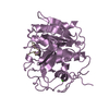
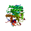
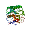
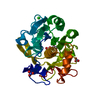
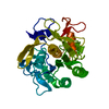
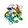
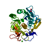
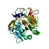
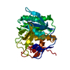
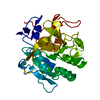
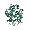
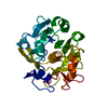
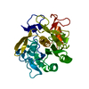
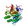
| Title | CRYSTAL STRUCTURE OF CALCIUM-FREE PROTEINASE K AT 1.5 ANGSTROMS RESOLUTION | ||||||
 Components Components | PROTEINASE K | ||||||
 Keywords Keywords | HYDROLASE(SERINE PROTEINASE) | ||||||
| Function / homology |  Function and homology information Function and homology informationpeptidase K / serine-type endopeptidase activity / proteolysis / extracellular region / metal ion binding Similarity search - Function | ||||||
| Biological species |  Engyodontium album (fungus) Engyodontium album (fungus) | ||||||
| Method |  X-RAY DIFFRACTION / Resolution: 1.5 Å X-RAY DIFFRACTION / Resolution: 1.5 Å | ||||||
 Authors Authors | Mueller, A. / Hinrichs, W. / Wolf, W.M. / Saenger, W. | ||||||
 Citation Citation | Journal: J.Biol.Chem. / Year: 1994 Title: Crystal structure of calcium-free proteinase K at 1.5-A resolution. Authors: Muller, A. / Hinrichs, W. / Wolf, W.M. / Saenger, W. #1: Journal: Nature / Year: 1989Title: Long-Range Structural Changes in Proteinase K Triggered by Calcium Ion Removal Authors: Bajorath, J. / Raghunathan, S. / Hinrichs, W. / Saenger, W. #2: Journal: Eur.J.Biochem. / Year: 1986Title: Three-Dimensional Structure of Proteinase K at 0.15-Nm Resolution Authors: Betzel, C. / Pal, G.P. / Saenger, W. | ||||||
| History |
|
- Structure visualization
| Structure viewer | Molecule: MolmilJmol/JSmol |
|---|
- Downloads & links
-Download
| PDBx/mmCIF format | 2pkc.cif.gz | 66.7 KB | Display | PDBx/mmCIF format |
|---|---|---|---|---|
| PDB format | pdb2pkc.ent.gz | 49.1 KB | Display | PDB format |
| PDBx/mmJSON format | 2pkc.json.gz | Tree view | PDBx/mmJSON format | |
| Others |  Other downloads Other downloads |
-Validation report
| Arichive directory | https://data.pdbj.org/pub/pdb/validation_reports/pk/2pkcftp://data.pdbj.org/pub/pdb/validation_reports/pk/2pkc | HTTPS FTP |
|---|
-Related structure data
| Similar structure data |
|---|


-Links
 PDBj
PDBj

- Assembly
| Deposited unit | 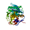
| ||||||||
|---|---|---|---|---|---|---|---|---|---|
| 1 |
| ||||||||
| Unit cell |
| ||||||||
| Atom site foot note | 1: CIS PROLINE - PRO 171 2: RESIDUES SER 15, SER 219, AND SER 247, SHOWED TWOFOLD DISORDERED POSITIONS FOR ATOM OG, HENCE THEIR ALTERNATE INDICATORS ARE *A* AND *B*. THEY HAVE AN OCCUPANCY OF 0.5 EACH, AND WERE REFINED AS DESCRIBED ABOVE. 3: ONE WATER (HOH 322) WAS LOCATED PRECISELY ON AN ELEMENT OF SYMMETRY, THEREFORE, IT WAS GIVEN HALF THE OCCUPANCY IT WOULD HAVE HAD, IF NOT LOCATED ON THE AXIS (OCC.=0.35). 4: ALL ATOMS OF THE PEPTIDE SEQUENCE WERE LOCATED, EXCEPT FOR THE SIDE-CHAIN OF THE C-TERMINAL GLN (278), WHICH WAS ONLY FOUND UP TO CB. | ||||||||
| Components on special symmetry positions |
|
-Components
| #1: Protein | Mass: 28930.783 Da / Num. of mol.: 1 Source method: isolated from a genetically manipulated source Source: (gene. exp.) Engyodontium album (fungus) / References: UniProt: P06873, peptidase K |
|---|---|
| #2: Chemical | ChemComp-NA /   Mass: 22.990 Da / Num. of mol.: 1 / Source method: obtained synthetically / Formula: Na Mass: 22.990 Da / Num. of mol.: 1 / Source method: obtained synthetically / Formula: Na |
| #3: Water | ChemComp-HOH /  Mass: 18.015 Da / Num. of mol.: 194 / Source method: isolated from a natural source / Formula: H2O Mass: 18.015 Da / Num. of mol.: 194 / Source method: isolated from a natural source / Formula: H2O |
| Compound details | THERE ARE THREE BENDS IN THIS STRUCTURE. S1: SER 79, GLY 83; S2: TRP 212, SER 216 (NESTED WITH T16); ...THERE ARE THREE BENDS IN THIS STRUCTURE. S1: SER 79, GLY 83; S2: TRP 212, SER 216 (NESTED WITH T16); S3: THR 244, ALA 248. |
| Has protein modification | Y |
| Sequence details | THE PROTEIN SEQUENCE (279 AMINO ACIDS) IS THE SAME AS THAT OF THE NATIVE ENZYME PRESENTED IN ...THE PROTEIN SEQUENCE (279 AMINO ACIDS) IS THE SAME AS THAT OF THE NATIVE ENZYME PRESENTED IN PROTEIN DATA BANK ENTRY 2PRK. |
-Experimental details
-Experiment
| Experiment | Method: X-RAY DIFFRACTION |
|---|
- Sample preparation
| Crystal | Density Matthews: 2.18 Å3/Da / Density % sol: 43.47 % | ||||||||||||||||||||||||||||||||||||
|---|---|---|---|---|---|---|---|---|---|---|---|---|---|---|---|---|---|---|---|---|---|---|---|---|---|---|---|---|---|---|---|---|---|---|---|---|---|
| Crystal grow | *PLUS pH: 7.5 / Method: vapor diffusion, sitting drop | ||||||||||||||||||||||||||||||||||||
| Components of the solutions | *PLUS
|
-Data collection
| Reflection | *PLUS Highest resolution: 1.5 Å / Lowest resolution: 10 Å / Num. obs: 33854 / % possible obs: 78 % / Observed criterion σ(I): 1 / Rmerge(I) obs: 0.147 |
|---|
- Processing
| Software | Name: TNT / Classification: refinement | ||||||||||||
|---|---|---|---|---|---|---|---|---|---|---|---|---|---|
| Refinement | Rfactor obs: 0.201 / Highest resolution: 1.5 Å Details: THE OCCUPANCIES OF WATER OXYGENS COULD BE REFINED IN A LAST CYCLE OF REFINEMENT, BUT ONLY THOSE WITH OCCUPANCY GREATER THAN 0.5 WERE RETAINED. | ||||||||||||
| Refinement step | Cycle: LAST / Highest resolution: 1.5 Å
| ||||||||||||
| Software | *PLUS Name: TNT / Classification: refinement | ||||||||||||
| Refinement | *PLUS Highest resolution: 1.5 Å / Rfactor obs: 0.201 | ||||||||||||
| Solvent computation | *PLUS | ||||||||||||
| Displacement parameters | *PLUS |
