 Movie
Movie Controller

[English] 日本語
 Yorodumi
- PDB-3de7: Proteinase K by Classical hanging drop method after the fourth st... -
+ Open data

- Basic information
| Entry | Database: PDB / ID: 3de7 | ||||||
|---|---|---|---|---|---|---|---|
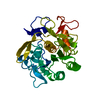
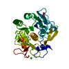
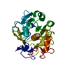
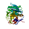
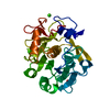
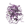
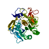
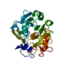
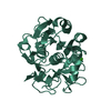
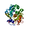
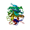
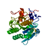
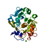
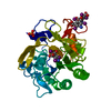
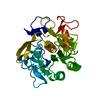
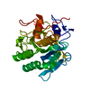
| Title | Proteinase K by Classical hanging drop method after the fourth step of high X-Ray dose on ESRF ID23-1 beamline | ||||||
 Components Components | Proteinase K | ||||||
 Keywords Keywords | HYDROLASE / Alpha Beta protein / Calcium / Metal-binding / Protease / Serine protease / Zymogen | ||||||
| Function / homology |  Function and homology information Function and homology informationpeptidase K / serine-type endopeptidase activity / proteolysis / extracellular region / metal ion binding Similarity search - Function | ||||||
| Biological species |  Engyodontium album (fungus) Engyodontium album (fungus) | ||||||
| Method |  X-RAY DIFFRACTION / SYNCHROTRON / MOLECULAR REPLACEMENT / Resolution: 2.3 Å X-RAY DIFFRACTION / SYNCHROTRON / MOLECULAR REPLACEMENT / Resolution: 2.3 Å | ||||||
 Authors Authors | Pechkova, E. / Tripathi, S.K. / Nicolini, C. | ||||||
 Citation Citation | Journal: J.Struct.Biol. / Year: 2009 Title: Radiation stability of proteinase K crystals grown by LB nanotemplate method Authors: Pechkova, E. / Tripathi, S. / Ravelli, R.B. / McSweeney, S. / Nicolini, C. | ||||||
| History |
|
- Structure visualization
| Structure viewer | Molecule: MolmilJmol/JSmol |
|---|
- Downloads & links
-Download
| PDBx/mmCIF format | 3de7.cif.gz | 68.1 KB | Display | PDBx/mmCIF format |
|---|---|---|---|---|
| PDB format | pdb3de7.ent.gz | 49.7 KB | Display | PDB format |
| PDBx/mmJSON format | 3de7.json.gz | Tree view | PDBx/mmJSON format | |
| Others |  Other downloads Other downloads |
-Validation report
| Arichive directory | https://data.pdbj.org/pub/pdb/validation_reports/de/3de7ftp://data.pdbj.org/pub/pdb/validation_reports/de/3de7 | HTTPS FTP |
|---|
-Related structure data
| Related structure data |  3d9qC  3ddzC  3de0C  3de1C  3de2C  3de3C  3de4C  3de5C  3de6C C: citing same article ( |
|---|---|
| Similar structure data |


-Links
 PDBj
PDBj

- Assembly
| Deposited unit | 
| ||||||||
|---|---|---|---|---|---|---|---|---|---|
| 1 |
| ||||||||
| Unit cell |
|
-Components
| #1: Protein | Mass: 28930.783 Da / Num. of mol.: 1 / Source method: isolated from a natural source / Source: (natural) Engyodontium album (fungus) / Strain: Limber / References: UniProt: P06873, peptidase K |
|---|---|
| #2: Chemical | ChemComp-CA /   Mass: 40.078 Da / Num. of mol.: 1 / Source method: obtained synthetically / Formula: Ca Mass: 40.078 Da / Num. of mol.: 1 / Source method: obtained synthetically / Formula: Ca |
| #3: Water | ChemComp-HOH /  Mass: 18.015 Da / Num. of mol.: 198 / Source method: isolated from a natural source / Formula: H2O Mass: 18.015 Da / Num. of mol.: 198 / Source method: isolated from a natural source / Formula: H2O |
| Has protein modification | Y |
-Experimental details
-Experiment
| Experiment | Method: X-RAY DIFFRACTION / Number of used crystals: 1 |
|---|
- Sample preparation
| Crystal | Density Matthews: 2.05 Å3/Da / Density % sol: 40.05 % / Mosaicity: 0.46 ° |
|---|---|
| Crystal grow | Temperature: 293 K / Method: vapor diffusion, hanging drop / pH: 7 Details: 20mg/ml of protein in 25mM HEPES, pH7.0, reservoir solution composed by 25mM HEPES and 400mM Na/K tartrate at pH7.0. Onto the siliconized glass cover slides were mixed 4 microL of protein ...Details: 20mg/ml of protein in 25mM HEPES, pH7.0, reservoir solution composed by 25mM HEPES and 400mM Na/K tartrate at pH7.0. Onto the siliconized glass cover slides were mixed 4 microL of protein solution with 4 microL of reservoir solution., VAPOR DIFFUSION, HANGING DROP, temperature 293K |
-Data collection
| Diffraction | Mean temperature: 100 K |
|---|---|
| Diffraction source | Source: SYNCHROTRON / Site: ESRF  / Beamline: ID23-1 / Wavelength: 0.97625 Å / Beamline: ID23-1 / Wavelength: 0.97625 Å |
| Detector | Type: ADSC QUANTUM 315 / Detector: CCD / Date: Feb 7, 2007 |
| Radiation | Monochromator: Si 111 CHANNEL / Protocol: SINGLE WAVELENGTH / Monochromatic (M) / Laue (L): M / Scattering type: x-ray |
| Radiation wavelength | Wavelength: 0.97625 Å / Relative weight: 1 |
| Reflection | Resolution: 2.3→56.705 Å / Num. obs: 11070 / % possible obs: 98.9 % / Redundancy: 4.6 % / Rmerge(I) obs: 0.173 / Rsym value: 0.173 / Net I/σ(I): 3.8 |
| Reflection shell | Resolution: 2.3→2.42 Å / Redundancy: 4.6 % / Rmerge(I) obs: 0.639 / Mean I/σ(I) obs: 1 / Num. measured all: 7489 / Num. unique all: 1614 / Rsym value: 0.639 / % possible all: 99.9 |
- Processing
| Software |
| |||||||||||||||||||||||||||||||||||||||||||||||||||||||||||||||||||||||||||||||||||||||||||||||
|---|---|---|---|---|---|---|---|---|---|---|---|---|---|---|---|---|---|---|---|---|---|---|---|---|---|---|---|---|---|---|---|---|---|---|---|---|---|---|---|---|---|---|---|---|---|---|---|---|---|---|---|---|---|---|---|---|---|---|---|---|---|---|---|---|---|---|---|---|---|---|---|---|---|---|---|---|---|---|---|---|---|---|---|---|---|---|---|---|---|---|---|---|---|---|---|---|
| Refinement | Method to determine structure: MOLECULAR REPLACEMENT / Resolution: 2.3→35.11 Å / Cor.coef. Fo:Fc: 0.945 / Cor.coef. Fo:Fc free: 0.924 / SU B: 5.988 / SU ML: 0.149 / Cross valid method: THROUGHOUT / σ(F): 0 / ESU R: 0.44 / ESU R Free: 0.228 / Stereochemistry target values: MAXIMUM LIKELIHOOD
| |||||||||||||||||||||||||||||||||||||||||||||||||||||||||||||||||||||||||||||||||||||||||||||||
| Solvent computation | Ion probe radii: 0.8 Å / Shrinkage radii: 0.8 Å / VDW probe radii: 1.2 Å / Solvent model: MASK | |||||||||||||||||||||||||||||||||||||||||||||||||||||||||||||||||||||||||||||||||||||||||||||||
| Displacement parameters | Biso mean: 15.784 Å2
| |||||||||||||||||||||||||||||||||||||||||||||||||||||||||||||||||||||||||||||||||||||||||||||||
| Refinement step | Cycle: LAST / Resolution: 2.3→35.11 Å
| |||||||||||||||||||||||||||||||||||||||||||||||||||||||||||||||||||||||||||||||||||||||||||||||
| Refine LS restraints |
| |||||||||||||||||||||||||||||||||||||||||||||||||||||||||||||||||||||||||||||||||||||||||||||||
| LS refinement shell | Resolution: 2.3→2.36 Å / Total num. of bins used: 20
|
