[English] 日本語
Yorodumi
- PDB-6zev: Crystal structure of proteinase K lamellae by electron diffractio... -

+
Open data


ID or keywords:

Loading...

-
Basic information

Entry
Database: PDB / ID: 6zev
TitleCrystal structure of proteinase K lamellae by electron diffraction with a 20 micrometre C2 condenser aperture
ComponentsProteinase K
KeywordsHYDROLASE / Protease / Serine protease
Function / homology
Function and homology information


peptidase K / serine-type endopeptidase activity / proteolysis / extracellular region / metal ion binding
Similarity search - Function
Proteinase K-like catalytic domain / Peptidase S8 propeptide/proteinase inhibitor I9 / Peptidase inhibitor I9 / Peptidase S8 propeptide/proteinase inhibitor I9 superfamily / Peptidase S8, subtilisin, His-active site / Serine proteases, subtilase family, histidine active site. / Serine proteases, subtilase family, aspartic acid active site. / Peptidase S8, subtilisin, Asp-active site / Serine proteases, subtilase family, serine active site. / Peptidase S8, subtilisin, Ser-active site ...Proteinase K-like catalytic domain / Peptidase S8 propeptide/proteinase inhibitor I9 / Peptidase inhibitor I9 / Peptidase S8 propeptide/proteinase inhibitor I9 superfamily / Peptidase S8, subtilisin, His-active site / Serine proteases, subtilase family, histidine active site. / Serine proteases, subtilase family, aspartic acid active site. / Peptidase S8, subtilisin, Asp-active site / Serine proteases, subtilase family, serine active site. / Peptidase S8, subtilisin, Ser-active site / Serine proteases, subtilase domain profile. / Peptidase S8, subtilisin-related / Peptidase S8/S53 domain superfamily / Subtilase family / Peptidase S8/S53 domain
Similarity search - Domain/homology
Biological speciesParengyodontium album (fungus)
MethodELECTRON CRYSTALLOGRAPHY / electron crystallography / MOLECULAR REPLACEMENT / cryo EM / Resolution: 2.4 Å
AuthorsEvans, G. / Zhang, P. / Beale, E.V. / Waterman, D.G.
Funding support United Kingdom, 3items
OrganizationGrant numberCountry
Wellcome Trust206422/Z/17/Z United Kingdom
Biotechnology and Biological Sciences Research Council (BBSRC)BB/S003339/1 United Kingdom
Wellcome Trust202933/Z/16/Z United Kingdom
Citation
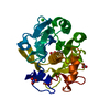
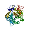
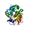
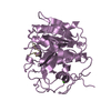
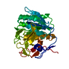
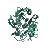
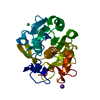
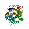
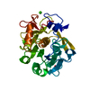
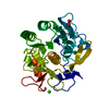
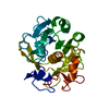
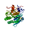
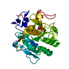
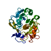
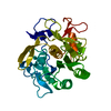
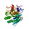
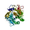
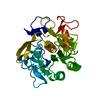
Journal: Front Mol Biosci / Year: 2020
Title: A Workflow for Protein Structure Determination From Thin Crystal Lamella by Micro-Electron Diffraction.
Authors: Emma V Beale / David G Waterman / Corey Hecksel / Jason van Rooyen / James B Gilchrist / James M Parkhurst / Felix de Haas / Bart Buijsse / Gwyndaf Evans / Peijun Zhang /
Abstract: MicroED has recently emerged as a powerful method for the analysis of biological structures at atomic resolution. This technique has been largely limited to protein nanocrystals which grow either as ...MicroED has recently emerged as a powerful method for the analysis of biological structures at atomic resolution. This technique has been largely limited to protein nanocrystals which grow either as needles or plates measuring only a few hundred nanometers in thickness. Furthermore, traditional microED data processing uses established X-ray crystallography software that is not optimized for handling compound effects that are unique to electron diffraction data. Here, we present an integrated workflow for microED, from sample preparation by cryo-focused ion beam milling, through data collection with a standard Ceta-D detector, to data processing using the DIALS software suite, thus enabling routine atomic structure determination of protein crystals of any size and shape using microED. We demonstrate the effectiveness of the workflow by determining the structure of proteinase K to 2.0 Å resolution and show the advantage of using protein crystal lamellae over nanocrystals.
History
DepositionJun 16, 2020Deposition site: PDBE / Processing site: PDBE
Revision 1.0Oct 14, 2020Provider: repository / Type: Initial release
Revision 1.1Nov 25, 2020Group: Structure summary / Category: em_entity_assembly
Revision 1.2Feb 10, 2021Group: Database references / Category: pdbx_related_exp_data_set
Revision 1.3Sep 13, 2023Group: Data collection / Database references ...Data collection / Database references / Derived calculations / Refinement description
Category: atom_type / chem_comp_atom ...atom_type / chem_comp_atom / chem_comp_bond / database_2 / pdbx_initial_refinement_model
Item: _atom_type.pdbx_N_electrons / _atom_type.pdbx_scat_Z ..._atom_type.pdbx_N_electrons / _atom_type.pdbx_scat_Z / _database_2.pdbx_DOI / _database_2.pdbx_database_accession
Revision 1.4Feb 14, 2024Group: Refinement description / Category: em_3d_fitting_list
Item: _em_3d_fitting_list.accession_code / _em_3d_fitting_list.initial_refinement_model_id ..._em_3d_fitting_list.accession_code / _em_3d_fitting_list.initial_refinement_model_id / _em_3d_fitting_list.source_name / _em_3d_fitting_list.type

-
Structure visualization

Movie
  • Deposited structure unit
  • Imaged by Jmol
  • Download
Movie viewer
Structure viewerMolecule:
MolmilJmol/JSmol

Downloads & links

-
Assembly

Deposited unit
AAA: Proteinase K
hetero molecules


Theoretical massNumber of molelcules
Total (without water)28,9992
Polymers28,9591
Non-polymers401
Water1086
1


  • Idetical with deposited unit
  • defined by author&software
TypeNameSymmetry operationNumber
identity operation1_555x,y,z1
Buried area90 Å2
ΔGint-12 kcal/mol
Surface area9960 Å2
MethodPISA
Unit cell
Length a, b, c (Å)67.328, 67.328, 106.599
Angle α, β, γ (deg.)90.000, 90.000, 90.000
Int Tables number96
Space group name H-MP43212

-
Components

#1: Protein Proteinase K / / Endopeptidase K / Tritirachium alkaline proteinase


Mass: 28958.791 Da / Num. of mol.: 1 / Source method: obtained synthetically / Details: Sigma-Aldrich, P2308 / Source: (synth.) Parengyodontium album (fungus) / References: UniProt: P06873, peptidase K
#2: Chemical ChemComp-CA / CALCIUM ION


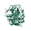
Mass: 40.078 Da / Num. of mol.: 1 / Source method: obtained synthetically / Formula: Ca
#3: Water ChemComp-HOH / water / Water


Mass: 18.015 Da / Num. of mol.: 6 / Source method: isolated from a natural source / Formula: H2O
Has ligand of interestN

-
Experimental details

-
Experiment

ExperimentMethod: ELECTRON CRYSTALLOGRAPHY
EM experimentAggregation state: 3D ARRAY / 3D reconstruction method: electron crystallography

-
Sample preparation

ComponentName: proteinase K / Type: COMPLEX / Source: MULTIPLE SOURCES
Buffer solutionpH: 7.5
Buffer componentConc.: 25 mM / Name: Tris
SpecimenConc.: 50 mg/ml / Embedding applied: NO / Shadowing applied: NO / Staining applied: NO / Vitrification applied: YES
Specimen supportGrid type: Quantifoil R1.2/1.3
VitrificationCryogen name: ETHANE / Humidity: 100 % / Chamber temperature: 293.15 K
Details: Excess liquid was removed by blotting for 4-6s with a Vitrobot

-
Data collection

Experimental equipment
Model: Talos Arctica / Image courtesy: FEI Company
MicroscopyModel: FEI TALOS ARCTICA / Date: Apr 18, 2018
Electron gunElectron source: FIELD EMISSION GUN / Accelerating voltage: 200 kV / Illumination mode: FLOOD BEAM
Electron lensMode: DIFFRACTION / C2 aperture diameter: 20 µm
Image recording

Imaging-ID: 1 / Average exposure time: 0.85 sec. / Electron dose: 0.034 e/Å2 / Film or detector model: OTHER / Num. of grids imaged: 1 / Details: The images were recorded using a Ceta-D detector operated in rolling-shutter mode with 2x2 binning.

IDNum. of diffraction images
144
278
345
4139
EM diffractionCamera length: 1350 mm
EM diffraction statsFourier space coverage: 100 % / High resolution: 2.4 Å / Num. of intensities measured: 121087 / Num. of structure factors: 10176 / Phase error: 0 ° / Phase residual: 0.1 ° / Phase error rejection criteria: none / Rmerge: 0.385 / Rsym: 0.385
Reflection shellResolution: 2.4→2.49 Å / Redundancy: 12.3 % / Rmerge(I) obs: 1.22 / Mean I/σ(I) obs: 2 / Num. measured obs: 12723 / Num. unique obs: 1035 / CC1/2: 0.701 / Rpim(I) all: 0.515 / Rrim(I) all: 1.327 / Χ2: 0.98 / % possible all: 99.7

-
Processing

Software
NameVersionClassification
REFMAC5.8.0258refinement
DIALS1.1data reduction
Aimless0.7.1data scaling
PHASER2.8.2phasing
EM software
IDNameVersionCategory
8REFMAC5.8.0258model refinement
9Phaser2.8.2molecular replacement
11POINTLESS1.11.12symmetry determination
12AIMLESS0.7.1crystallography merging
EM 3D crystal entity∠α: 90 ° / ∠β: 90 ° / ∠γ: 90 ° / A: 67.3277 Å / B: 67.3277 Å / C: 106.599 Å / Space group name: P43212 / Space group num: 96
CTF correctionType: NONE
3D reconstructionResolution method: DIFFRACTION PATTERN/LAYERLINES / Symmetry type: 3D CRYSTAL
Atomic model buildingSpace: RECIPROCAL
Atomic model buildingPDB-ID: 2ID8
Accession code: 2ID8 / Source name: PDB / Type: experimental model
RefinementMethod to determine structure: MOLECULAR REPLACEMENT
Starting model: 2ID8
Resolution: 2.4→56.989 Å / Cor.coef. Fo:Fc: 0.921 / Cor.coef. Fo:Fc free: 0.887 / Cross valid method: FREE R-VALUE / ESU R: 0.597 / ESU R Free: 0.274
RfactorNum. reflection% reflection
Rfree0.2427 495 4.897 %
Rwork0.1996 9614 -
all0.202 --
obs-10109 99.881 %
Solvent computationIon probe radii: 0.8 Å / Shrinkage radii: 0.8 Å / VDW probe radii: 1.2 Å / Solvent model: MASK BULK SOLVENT
Displacement parametersBiso mean: 18.302 Å2
Baniso -1Baniso -2Baniso -3
1--1.041 Å20 Å20 Å2
2---1.041 Å20 Å2
3---2.082 Å2
Refine LS restraints
Refine-IDTypeDev idealDev ideal targetNumber
ELECTRON CRYSTALLOGRAPHYr_bond_refined_d0.0070.0122098
ELECTRON CRYSTALLOGRAPHYr_angle_refined_deg1.241.6322858
ELECTRON CRYSTALLOGRAPHYr_dihedral_angle_1_deg5.2455285
ELECTRON CRYSTALLOGRAPHYr_dihedral_angle_2_deg35.85322.2100
ELECTRON CRYSTALLOGRAPHYr_dihedral_angle_3_deg13.74715306
ELECTRON CRYSTALLOGRAPHYr_dihedral_angle_4_deg18.8371512
ELECTRON CRYSTALLOGRAPHYr_chiral_restr0.0530.2282
ELECTRON CRYSTALLOGRAPHYr_gen_planes_refined0.0030.021646
ELECTRON CRYSTALLOGRAPHYr_nbd_refined0.2060.21075
ELECTRON CRYSTALLOGRAPHYr_nbtor_refined0.3190.21475
ELECTRON CRYSTALLOGRAPHYr_xyhbond_nbd_refined0.1350.281
ELECTRON CRYSTALLOGRAPHYr_metal_ion_refined0.0110.21
ELECTRON CRYSTALLOGRAPHYr_symmetry_nbd_refined0.2770.240
ELECTRON CRYSTALLOGRAPHYr_symmetry_xyhbond_nbd_refined0.2470.24
ELECTRON CRYSTALLOGRAPHYr_mcbond_it4.8671.541119
ELECTRON CRYSTALLOGRAPHYr_mcangle_it5.872.3161399
ELECTRON CRYSTALLOGRAPHYr_scbond_it7.2122.051979
ELECTRON CRYSTALLOGRAPHYr_scangle_it7.8682.8161455
ELECTRON CRYSTALLOGRAPHYr_lrange_it8.62122.7763327
LS refinement shell
Resolution (Å)Rfactor RfreeNum. reflection RfreeRfactor RworkNum. reflection RworkRefine-ID% reflection obs (%)
2.4-2.4630.311310.275693ELECTRON CRYSTALLOGRAPHY100
2.463-2.530.253320.252674ELECTRON CRYSTALLOGRAPHY99.7175
2.53-2.6030.293280.249673ELECTRON CRYSTALLOGRAPHY100
2.603-2.6830.376390.229628ELECTRON CRYSTALLOGRAPHY99.8503
2.683-2.7710.367260.22614ELECTRON CRYSTALLOGRAPHY100
2.771-2.8690.322340.204613ELECTRON CRYSTALLOGRAPHY99.8457
2.869-2.9770.239330.205578ELECTRON CRYSTALLOGRAPHY99.8366
2.977-3.0980.306240.194557ELECTRON CRYSTALLOGRAPHY99.8282
3.098-3.2360.22280.176557ELECTRON CRYSTALLOGRAPHY100
3.236-3.3940.236320.172508ELECTRON CRYSTALLOGRAPHY100
3.394-3.5770.231230.161497ELECTRON CRYSTALLOGRAPHY100
3.577-3.7940.127280.147461ELECTRON CRYSTALLOGRAPHY100
3.794-4.0550.158230.142437ELECTRON CRYSTALLOGRAPHY100
4.055-4.380.163240.143425ELECTRON CRYSTALLOGRAPHY100
4.38-4.7970.197230.138388ELECTRON CRYSTALLOGRAPHY99.5157
4.797-5.3620.15240.175345ELECTRON CRYSTALLOGRAPHY100
5.362-6.1890.165160.217322ELECTRON CRYSTALLOGRAPHY99.4118
6.189-7.5730.378140.218277ELECTRON CRYSTALLOGRAPHY100
7.573-10.6830.2780.258224ELECTRON CRYSTALLOGRAPHY99.5708
10.683-56.9890.86950.553143ELECTRON CRYSTALLOGRAPHY99.3289

+
About Yorodumi

-
News

-
Feb 9, 2022. New format data for meta-information of EMDB entries

New format data for meta-information of EMDB entries

  • Version 3 of the EMDB header file is now the official format.
  • The previous official version 1.9 will be removed from the archive.

Related info.:EMDB header

External links:wwPDB to switch to version 3 of the EMDB data model

-
Aug 12, 2020. Covid-19 info

Covid-19 info

URL: https://pdbj.org/emnavi/covid19.php

New page: Covid-19 featured information page in EM Navigator.

Related info.:Covid-19 info / Mar 5, 2020. Novel coronavirus structure data

+
Mar 5, 2020. Novel coronavirus structure data

Novel coronavirus structure data

Related info.:Yorodumi Speices / Aug 12, 2020. Covid-19 info

External links:COVID-19 featured content - PDBj / Molecule of the Month (242):Coronavirus Proteases

+
Jan 31, 2019. EMDB accession codes are about to change! (news from PDBe EMDB page)

EMDB accession codes are about to change! (news from PDBe EMDB page)

  • The allocation of 4 digits for EMDB accession codes will soon come to an end. Whilst these codes will remain in use, new EMDB accession codes will include an additional digit and will expand incrementally as the available range of codes is exhausted. The current 4-digit format prefixed with “EMD-” (i.e. EMD-XXXX) will advance to a 5-digit format (i.e. EMD-XXXXX), and so on. It is currently estimated that the 4-digit codes will be depleted around Spring 2019, at which point the 5-digit format will come into force.
  • The EM Navigator/Yorodumi systems omit the EMD- prefix.

Related info.:Q: What is EMD? / ID/Accession-code notation in Yorodumi/EM Navigator

External links:EMDB Accession Codes are Changing Soon! / Contact to PDBj

+
Jul 12, 2017. Major update of PDB

Major update of PDB

  • wwPDB released updated PDB data conforming to the new PDBx/mmCIF dictionary.
  • This is a major update changing the version number from 4 to 5, and with Remediation, in which all the entries are updated.
  • In this update, many items about electron microscopy experimental information are reorganized (e.g. em_software).
  • Now, EM Navigator and Yorodumi are based on the updated data.

External links:wwPDB Remediation / Enriched Model Files Conforming to OneDep Data Standards Now Available in the PDB FTP Archive

-
Yorodumi

Thousand views of thousand structures

  • Yorodumi is a browser for structure data from EMDB, PDB, SASBDB, etc.
  • This page is also the successor to EM Navigator detail page, and also detail information page/front-end page for Omokage search.
  • The word "yorodu" (or yorozu) is an old Japanese word meaning "ten thousand". "mi" (miru) is to see.

Related info.:EMDB / PDB / SASBDB / Comparison of 3 databanks / Yorodumi Search / Aug 31, 2016. New EM Navigator & Yorodumi / Yorodumi Papers / Jmol/JSmol / Function and homology information / Changes in new EM Navigator and Yorodumi

Read more