 ムービー
ムービー コントローラー
 構造ビューア
 EMN検索について


-検索条件
-検索結果
検索 (著者・登録者: luo & d)の結果635件中、1から50件目までを表示しています
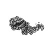
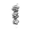
EMDB-38763: 
Fab M2-7 complexed with SARS-Cov2 RBD and human ACE2
PDB-8xxw:
Fab M2-7 complexed with SARS-Cov2 RBD and human ACE2

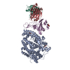
EMDB-37640:
Cryo-EM structure of DiCas7-11 in complex with crRNA

EMDB-37649:
Cryo-EM structure of DiCas7-11-crRNA in complex with regulator

EMDB-37653:
Cryo-EM structure of DiCas7-11 mutant in complex with crRNA

EMDB-37655:
Cryo-EM structure of Cas7-11-crRNA bound to N-terminal of TPR-CHAT

PDB-8wm4:
Cryo-EM structure of DiCas7-11 in complex with crRNA

PDB-8wmc:
Cryo-EM structure of DiCas7-11-crRNA in complex with regulator

PDB-8wmi:
Cryo-EM structure of DiCas7-11 mutant in complex with crRNA

PDB-8wml:
Cryo-EM structure of Cas7-11-crRNA bound to N-terminal of TPR-CHAT

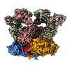
EMDB-17356:
Structure of divisome complex FtsWIQLB

EMDB-38906:
Cell divisome sPG hydrolysis machinery FtsEX-EnvC

PDB-8y3x:
Cell divisome sPG hydrolysis machinery FtsEX-EnvC
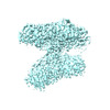
EMDB-37663:
CryoEM structure of ZIKV rsNS1
EMDB-37670:
ZIKV rsNS1 in complex with Fab AA12 and anti-fab nanobody

EMDB-37673:
ZIKV rsNS1 in complex with Fab GB5 and anti-fab nanobody
EMDB-37676:
CryoEM structure of ZIKV rsNS1 filament
EMDB-37678:
ZIKV rsNS1 in complex with Fab EB9 and anti-fab nanobody
PDB-8wn8:
CryoEM structure of ZIKV rsNS1
PDB-8wnp:
ZIKV rsNS1 in complex with Fab AA12 and anti-fab nanobody
PDB-8wnu:
ZIKV rsNS1 in complex with Fab GB5 and anti-fab nanobody

PDB-8wo0:
CryoEM structure of ZIKV rsNS1 filament
PDB-8wo4:
ZIKV rsNS1 in complex with Fab EB9 and anti-fab nanobody

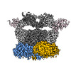
EMDB-38453:
Structure of the SARS-CoV-2 EG.5.1 spike glycoprotein in complex with ACE2 (1-up state)

EMDB-38454:
Structure of the SARS-CoV-2 EG.5.1 spike RBD in complex with ACE2

PDB-8xlm:
Structure of the SARS-CoV-2 EG.5.1 spike glycoprotein in complex with ACE2 (1-up state)

PDB-8xln:
Structure of the SARS-CoV-2 EG.5.1 spike RBD in complex with ACE2

EMDB-37648:
SARS-CoV-2 EG.5.1 spike glycoprotein (1-up state)

EMDB-37650:
SARS-CoV-2 EG.5.1 spike glycoprotein (closed-2 state)

EMDB-37651:
SARS-CoV-2 EG.5.1 spike glycoprotein (closed-1 state)

PDB-8wmd:
Structure of the SARS-CoV-2 EG.5.1 spike glycoprotein (closed-2 state)

PDB-8wmf:
Structure of the SARS-CoV-2 EG.5.1 spike glycoprotein (closed-1 state)

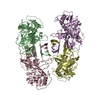
EMDB-36800:
Potassium transporter KtrAB from Bacillus subtilis in ADP-bound state

EMDB-36801:
Potassium transporter KtrAB from Bacillus subtilis in ADP-bound state, focused refined on KtrA octamer

EMDB-36802:
Potassium transporter KtrAB from Bacillus subtilis in ADP-bound state, focused refined on KtrB dimer

EMDB-36803:
Potassium transporter KtrAB from Bacillus subtilis in ATP-bound state with addition of MgCl2

EMDB-36804:
Potassium transporter KtrAB from Bacillus subtilis in ATP-bound state with addition of EDTA and EGTA

EMDB-38477:
Potassium transporter KtrAB from Bacillus subtilis in ATP-bound state with addition of EDTA and EGTA, vertical C2 symmetry axis

EMDB-38478:
Potassium transporter KtrAB from Bacillus subtilis in ATP-bound state with addition of EDTA and EGTA, C1 symmetry

PDB-8k1s:
Potassium transporter KtrAB from Bacillus subtilis in ADP-bound state

PDB-8k1t:
Potassium transporter KtrAB from Bacillus subtilis in ATP-bound state with addition of MgCl2

PDB-8k1u:
Potassium transporter KtrAB from Bacillus subtilis in ATP-bound state with addition of EDTA and EGTA

PDB-8xmh:
Potassium transporter KtrAB from Bacillus subtilis in ATP-bound state with addition of EDTA and EGTA, vertical C2 symmetry axis

PDB-8xmi:
Potassium transporter KtrAB from Bacillus subtilis in ATP-bound state with addition of EDTA and EGTA, C1 symmetry

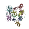
EMDB-37690:
Structure of the wild-type Arabidopsis ABCB19 in the apo state

EMDB-37692:
Structure of the wild-type Arabidopsis ABCB19 in the brassinolide-bound state

EMDB-37694:
Structure of the wild-type Arabidopsis ABCB19 in the brassinolide and AMP-PNP bound state

EMDB-37705:
Structure of the Arabidopsis E529Q/E1174Q ABCB19 in the ATP bound state

PDB-8woi:
Structure of the wild-type Arabidopsis ABCB19 in the apo state

PDB-8wom:
Structure of the wild-type Arabidopsis ABCB19 in the brassinolide-bound state
ページ:
 wwPDBはEMDBデータモデルのバージョン3へ移行します
