+ Open data
- Basic information
| Entry | Database: PDB / ID: 8y3x | |||||||||||||||||||||||||||||||||||||||||||||
|---|---|---|---|---|---|---|---|---|---|---|---|---|---|---|---|---|---|---|---|---|---|---|---|---|---|---|---|---|---|---|---|---|---|---|---|---|---|---|---|---|---|---|---|---|---|---|
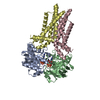
| Title | Cell divisome sPG hydrolysis machinery FtsEX-EnvC | |||||||||||||||||||||||||||||||||||||||||||||
 Components Components |
| |||||||||||||||||||||||||||||||||||||||||||||
 Keywords Keywords | MEMBRANE PROTEIN / CELL DIVISION | |||||||||||||||||||||||||||||||||||||||||||||
| Function / homology |  Function and homology information Function and homology informationdivision septum / divisome complex / peptidoglycan-based cell wall biogenesis / Gram-negative-bacterium-type cell wall / septum digestion after cytokinesis / peptidoglycan turnover / plasma membrane protein complex / division septum assembly / FtsZ-dependent cytokinesis / extrinsic component of membrane ...division septum / divisome complex / peptidoglycan-based cell wall biogenesis / Gram-negative-bacterium-type cell wall / septum digestion after cytokinesis / peptidoglycan turnover / plasma membrane protein complex / division septum assembly / FtsZ-dependent cytokinesis / extrinsic component of membrane / cell division site / ATPase complex / positive regulation of cell division / transmembrane transporter activity / response to radiation / metalloendopeptidase activity / transmembrane transport / outer membrane-bounded periplasmic space / periplasmic space / hydrolase activity / response to xenobiotic stimulus / cell division / response to antibiotic / ATP hydrolysis activity / ATP binding / membrane / plasma membrane / cytoplasm Similarity search - Function | |||||||||||||||||||||||||||||||||||||||||||||
| Biological species |  | |||||||||||||||||||||||||||||||||||||||||||||
| Method | ELECTRON MICROSCOPY / single particle reconstruction / cryo EM / Resolution: 3.11 Å | |||||||||||||||||||||||||||||||||||||||||||||
 Authors Authors | Zhang, Z. / Dong, H. / Chen, Y. | |||||||||||||||||||||||||||||||||||||||||||||
| Funding support |  China, 1items China, 1items
| |||||||||||||||||||||||||||||||||||||||||||||
 Citation Citation |  Journal: PLoS Biol / Year: 2024 Journal: PLoS Biol / Year: 2024Title: Structure and activity of the septal peptidoglycan hydrolysis machinery crucial for bacterial cell division. Authors: Yatian Chen / Jiayue Gu / Biao Yang / Lili Yang / Jie Pang / Qinghua Luo / Yirong Li / Danyang Li / Zixin Deng / Changjiang Dong / Haohao Dong / Zhengyu Zhang /  Abstract: The peptidoglycan (PG) layer is a critical component of the bacterial cell wall and serves as an important target for antibiotics in both gram-negative and gram-positive bacteria. The hydrolysis of ...The peptidoglycan (PG) layer is a critical component of the bacterial cell wall and serves as an important target for antibiotics in both gram-negative and gram-positive bacteria. The hydrolysis of septal PG (sPG) is a crucial step of bacterial cell division, facilitated by FtsEX through an amidase activation system. In this study, we present the cryo-EM structures of Escherichia coli FtsEX and FtsEX-EnvC in the ATP-bound state at resolutions of 3.05 Å and 3.11 Å, respectively. Our PG degradation assays in E. coli reveal that the ATP-bound conformation of FtsEX activates sPG hydrolysis of EnvC-AmiB, whereas EnvC-AmiB alone exhibits autoinhibition. Structural analyses indicate that ATP binding induces conformational changes in FtsEX-EnvC, leading to significant differences from the apo state. Furthermore, PG degradation assays of AmiB mutants confirm that the regulation of AmiB by FtsEX-EnvC is achieved through the interaction between EnvC-AmiB. These findings not only provide structural insight into the mechanism of sPG hydrolysis and bacterial cell division, but also have implications for the development of novel therapeutics targeting drug-resistant bacteria. | |||||||||||||||||||||||||||||||||||||||||||||
| History |
|
- Structure visualization
| Structure viewer | Molecule:  Molmil Molmil Jmol/JSmol Jmol/JSmol |
|---|
- Downloads & links
- Download
| PDBx/mmCIF format |  8y3x.cif.gz 8y3x.cif.gz | 218.4 KB | Display |  PDBx/mmCIF format PDBx/mmCIF format |
|---|---|---|---|---|
| PDB format |  pdb8y3x.ent.gz pdb8y3x.ent.gz | 161.3 KB | Display |  PDB format PDB format |
| PDBx/mmJSON format |  8y3x.json.gz 8y3x.json.gz | Tree view |  PDBx/mmJSON format PDBx/mmJSON format | |
| Others |  Other downloads Other downloads |
-Validation report
| Arichive directory |  https://data.pdbj.org/pub/pdb/validation_reports/y3/8y3x https://data.pdbj.org/pub/pdb/validation_reports/y3/8y3x ftp://data.pdbj.org/pub/pdb/validation_reports/y3/8y3x ftp://data.pdbj.org/pub/pdb/validation_reports/y3/8y3x | HTTPS FTP |
|---|
-Related structure data
| Related structure data |  38906MC  8x61C M: map data used to model this data C: citing same article ( |
|---|---|
| Similar structure data | Similarity search - Function & homology  F&H Search F&H Search |
- Links
Links
- Assembly
| Deposited unit | 
|
|---|---|
| 1 |
|
- Components
| #1: Protein | Mass: 24475.295 Da / Num. of mol.: 2 / Mutation: E163Q Source method: isolated from a genetically manipulated source Source: (gene. exp.)   #2: Protein | Mass: 38583.500 Da / Num. of mol.: 2 Source method: isolated from a genetically manipulated source Source: (gene. exp.)   #3: Protein | | Mass: 46661.617 Da / Num. of mol.: 1 Source method: isolated from a genetically manipulated source Source: (gene. exp.)   #4: Chemical | Has ligand of interest | Y | Has protein modification | N | |
|---|
-Experimental details
-Experiment
| Experiment | Method: ELECTRON MICROSCOPY |
|---|---|
| EM experiment | Aggregation state: PARTICLE / 3D reconstruction method: single particle reconstruction |
- Sample preparation
| Component | Name: FtsEX-EnvC / Type: COMPLEX / Entity ID: #1-#3 / Source: RECOMBINANT |
|---|---|
| Source (natural) | Organism:  |
| Source (recombinant) | Organism:  |
| Buffer solution | pH: 8 |
| Specimen | Conc.: 1 mg/ml / Embedding applied: NO / Shadowing applied: NO / Staining applied: NO / Vitrification applied: YES |
| Vitrification | Cryogen name: ETHANE / Humidity: 100 % |
- Electron microscopy imaging
| Experimental equipment |  Model: Titan Krios / Image courtesy: FEI Company |
|---|---|
| Microscopy | Model: FEI TITAN KRIOS |
| Electron gun | Electron source:  FIELD EMISSION GUN / Accelerating voltage: 300 kV / Illumination mode: OTHER FIELD EMISSION GUN / Accelerating voltage: 300 kV / Illumination mode: OTHER |
| Electron lens | Mode: OTHER / Nominal defocus max: 3000 nm / Nominal defocus min: 1000 nm |
| Image recording | Electron dose: 60 e/Å2 / Film or detector model: GATAN K3 BIOQUANTUM (6k x 4k) |
- Processing
| EM software | Name: PHENIX / Category: model refinement | ||||||||||||||||||||||||
|---|---|---|---|---|---|---|---|---|---|---|---|---|---|---|---|---|---|---|---|---|---|---|---|---|---|
| CTF correction | Type: NONE | ||||||||||||||||||||||||
| 3D reconstruction | Resolution: 3.11 Å / Resolution method: FSC 0.143 CUT-OFF / Num. of particles: 4039317 / Symmetry type: POINT | ||||||||||||||||||||||||
| Refine LS restraints |
|
 Movie
Movie Controller


 PDBj
PDBj


