 ムービー
ムービー コントローラー
 構造ビューア
 EMN検索について


-検索条件
-検索結果
検索 (著者・登録者: galkin & ve)の結果全42件を表示しています
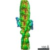
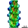
EMDB-42681: 
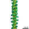
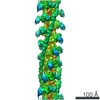
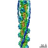
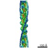
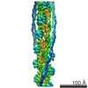
The structure of the native cardiac thin filament troponin core in Ca2+-free state from the upper strand
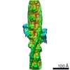
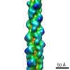
EMDB-42682:
The structure of the native cardiac thin filament troponin core in Ca2+-free tilted state from the upper strand

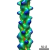
EMDB-42683:
The structure of the native cardiac thin filament troponin core in Ca2+-free rotated state from the upper strand
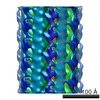
EMDB-42800:
The structure of the native cardiac thin filament troponin core in Ca2+-free state from the lower strand
EMDB-42833:
The structure of the native cardiac thin filament troponin core in Ca2+-free rotated state from the lower strand
EMDB-42835:
The structure of the native cardiac thin filament troponin core in Ca2+-free tilted state from the lower strand
EMDB-42846:
The structure of the native cardiac thin filament troponin core in Ca2+-bound fully activated state from the upper strand

EMDB-42847:
The structure of the native cardiac thin filament troponin core in Ca2+-bound partially activated state from the upper strand
EMDB-42849:
The structure of the native cardiac thin filament troponin core in Ca2+-bound fully activated state 1 from the lower strand
EMDB-42856:
The structure of the native cardiac thin filament troponin core in Ca2+-bound fully activated state 2 from the lower strand
EMDB-42858:
The structure of the native cardiac thin filament troponin core in Ca2+-bound partially activated state from the lower strand

EMDB-42874:
The structure of the native cardiac thin filament troponin core in Ca2+-free state from the upper strand activated by the C1-domain of cardiac myosin binding protein C
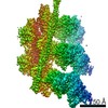
EMDB-27331:
The structure of the native cardiac thin filament junction region

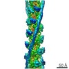
EMDB-25911:
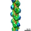
Cardiac F-actin decorated with regulatory M-domain of cardiac myosin binding protein C

EMDB-25914:
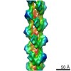
Cardiac thin filament decorated with regulatory M-domain of cardiac myosin binding protein C

EMDB-25918:
Cardiac thin filament decorated with C1 Ig-domain and regulatory M-domain of cardiac myosin binding protein C (cMyBP-C)

EMDB-23497:
Filamentous actin decorated with human cardiac myosin binding protein C C2 domain

EMDB-22964:
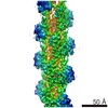
Structure of cardiac native thin filament at pCa=5.8 having upper and lower troponins in Ca2+ free state
EMDB-22965:
Structure of cardiac native thin filament at pCa=5.8 having upper and lower troponins in Ca2+ bound state
EMDB-22966:
Structure of the native cardiac thin filament at pCa=5.8 having upper Tn in Ca2+ free state and lower Tn in Ca2+ bound state
EMDB-22978:
Structure of upper Tn Ca2+ free (rotated) and lower Tn Ca2+ bound cardiac native thin filament at pCa=5.8
EMDB-22981:
Structure of cardiac native thin filament at pCa=5.8 having upper troponin in Ca2+ bound state and lower troponin in Ca2+ free state
EMDB-22335:
cardiac actomyosin complex
EMDB-4346:
human cardiac myosin binding protein C C1 Ig-domain bound to native cardiac thin filament
EMDB-7780:
Cardiac thin filament decorated with C0C1 fragment of cardiac myosin binding protein C mode 1
EMDB-7781:
Cardiac thin filament decorated with C0C1 fragment of cardiac myosin binding protein C mode 2
EMDB-3666:
Ca2+-induced Movement of Tropomyosin on Native Cardiac Thin Filaments - "OPEN" state
EMDB-3665:
Ca2+-induced Movement of Tropomyosin on Native Cardiac Thin Filaments - "Blocked" state
EMDB-3667:
Ca2+-induced Movement of Tropomyosin on Native Cardiac Thin Filaments - "Closed" state
EMDB-6179:
One state of F-Actin at near-atomic resolution
EMDB-6180:
Tilted state of actin, T1
EMDB-6181:
Tilted state of actin, T2
EMDB-1951:
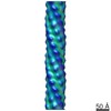
Structure of the CS1 pilus of enterotoxigenic Escherichia coli reveals structural polymorphism
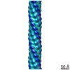
EMDB-1952:
Structure of the CS1 pilus of enterotoxigenic Escherichia coli reveals structural polymorphism
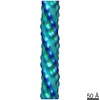
EMDB-1953:
Structure of the CS1 pilus of enterotoxigenic Escherichia coli reveals structural polymorphism
EMDB-5354:
Remodeling of actin filaments by ADF-cofilin proteins
EMDB-5170:
Binding of alpha-actinin CH1 to F-actin

PDB-3iku:
Structural model of ParM filament in closed state from cryo-EM

PDB-3iky:
Structural model of ParM filament in the open state by cryo-EM
EMDB-5128:
Reconstruction of ParM in the closed state using cryo-EM
EMDB-5129:
Reconstruction of ParM in the open state using cryo-EM
EMDB-5007:
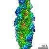
Divergence of Quaternary Structures among Bacterial Flagellar Filaments
 wwPDBはEMDBデータモデルのバージョン3へ移行します
