 Movie
Movie Controller

[English] 日本語
 Yorodumi
- EMDB-5007: Divergence of Quaternary Structures among Bacterial Flagellar Fil... -
+ Open data

- Basic information
| Entry | Database: EMDB / ID: EMD-5007 | |||||||||
|---|---|---|---|---|---|---|---|---|---|---|
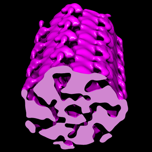
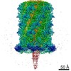
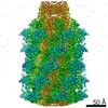
| Title | Divergence of Quaternary Structures among Bacterial Flagellar Filaments | |||||||||
 Map data Map data | Volume | |||||||||
 Sample Sample |
| |||||||||
 Keywords Keywords | flagellar filament / structural polymorphism / IHRSR | |||||||||
| Biological species |   Campylobacter jejuni (Campylobacter) Campylobacter jejuni (Campylobacter) | |||||||||
| Method | helical reconstruction / cryo EM | |||||||||
 Authors Authors | Galkin VE / Yu X / Bielnicki J / Heuser J / Ewing CP / Guerry P / Egelman EH | |||||||||
 Citation Citation | Journal: Science / Year: 2008 Title: Divergence of quaternary structures among bacterial flagellar filaments. Authors: Vitold E Galkin / Xiong Yu / Jakub Bielnicki / John Heuser / Cheryl P Ewing / Patricia Guerry / Edward H Egelman /  Abstract: It has been widely assumed that the atomic structure of the flagellar filament from Salmonella typhimurium serves as a model for all bacterial flagellar filaments given the sequence conservation in ...It has been widely assumed that the atomic structure of the flagellar filament from Salmonella typhimurium serves as a model for all bacterial flagellar filaments given the sequence conservation in the coiled-coil regions responsible for polymerization. On the basis of electron microscopic images, we show that the flagellar filaments from Campylobacter jejuni have seven protofilaments rather than the 11 in S. typhimurium. The vertebrate Toll-like receptor 5 (TLR5) recognizes a region of bacterial flagellin that is involved in subunit-subunit assembly in Salmonella and many other pathogenic bacteria, and this short region has diverged in Campylobacter and related bacteria, such as Helicobacter pylori, which are not recognized by TLR5. The driving force in the change of quaternary structure between Salmonella and Campylobacter may have been the evasion of TLR5. | |||||||||
| History |
|
- Structure visualization
| Movie |
 Movie viewer Movie viewer |
|---|---|
| Structure viewer | EM map: SurfViewMolmilJmol/JSmol |
| Supplemental images |
- Downloads & links
-EMDB archive
| Map data | emd_5007.map.gz | 3.5 MB | EMDB map data format | |
|---|---|---|---|---|
| Header (meta data) | emd-5007-v30.xmlemd-5007.xml | 6.8 KB 6.8 KB | Display Display | EMDB header |
| Images |  emd_5007_1.png emd_5007_1.png | 123.3 KB | ||
| Archive directory |  http://ftp.pdbj.org/pub/emdb/structures/EMD-5007ftp://ftp.pdbj.org/pub/emdb/structures/EMD-5007 http://ftp.pdbj.org/pub/emdb/structures/EMD-5007ftp://ftp.pdbj.org/pub/emdb/structures/EMD-5007 | HTTPS FTP |
-Related structure data
| Similar structure data |
|---|
-Links
| EMDB pages | EMDB (EBI/PDBe) / EMDataResource |
|---|
-Map
| File | Download / File: emd_5007.map.gz / Format: CCP4 / Size: 3.7 MB / Type: IMAGE STORED AS FLOATING POINT NUMBER (4 BYTES) | ||||||||||||||||||||||||||||||||||||||||||||||||||||||||||||
|---|---|---|---|---|---|---|---|---|---|---|---|---|---|---|---|---|---|---|---|---|---|---|---|---|---|---|---|---|---|---|---|---|---|---|---|---|---|---|---|---|---|---|---|---|---|---|---|---|---|---|---|---|---|---|---|---|---|---|---|---|---|
| Annotation | Volume | ||||||||||||||||||||||||||||||||||||||||||||||||||||||||||||
| Projections & slices | Image control
Images are generated by Spider. | ||||||||||||||||||||||||||||||||||||||||||||||||||||||||||||
| Voxel size | X=Y=Z: 2.4 Å | ||||||||||||||||||||||||||||||||||||||||||||||||||||||||||||
| Density |
| ||||||||||||||||||||||||||||||||||||||||||||||||||||||||||||
| Symmetry | Space group: 1 | ||||||||||||||||||||||||||||||||||||||||||||||||||||||||||||
| Details | EMDB XML:
CCP4 map header:
| ||||||||||||||||||||||||||||||||||||||||||||||||||||||||||||
 Z (Sec.)
 Y (Row.)
 X (Col.)


-Supplemental data
- Sample components
-Entire : Campylobacter jejuni flagellar filament
| Entire | Name: Campylobacter jejuni flagellar filament |
|---|---|
| Components |
|
-Supramolecule #1000: Campylobacter jejuni flagellar filament
| Supramolecule | Name: Campylobacter jejuni flagellar filament / type: sample / ID: 1000 / Details: None / Oligomeric state: helical polymer / Number unique components: 1 |
|---|
-Supramolecule #1: flagellar filament
| Supramolecule | Name: flagellar filament / type: organelle_or_cellular_component / ID: 1 / Recombinant expression: No / Database: NCBI |
|---|---|
| Source (natural) | Organism: Campylobacter jejuni (Campylobacter) / Organelle: Flagella |
-Experimental details
-Structure determination
| Method | cryo EM |
|---|---|
 Processing Processing | helical reconstruction |
| Aggregation state | filament |
-Sample preparation
| Vitrification | Cryogen name: ETHANE / Instrument: OTHER |
|---|
- Electron microscopy
| Microscope | FEI TECNAI F20 |
|---|---|
| Electron beam | Acceleration voltage: 200 kV / Electron source:  FIELD EMISSION GUN FIELD EMISSION GUN |
| Electron optics | Illumination mode: OTHER / Imaging mode: BRIGHT FIELD |
| Sample stage | Specimen holder: Eucentric / Specimen holder model: GATAN LIQUID NITROGEN |
| Experimental equipment |  Model: Tecnai F20 / Image courtesy: FEI Company |
-Image processing
| Final reconstruction | Applied symmetry - Helical parameters - Δz: 7.5 Å Applied symmetry - Helical parameters - Δ&Phi: 103.11 ° Applied symmetry - Helical parameters - Axial symmetry: C1 (asymmetric) Algorithm: OTHER |
|---|
