 Movie
Movie Controller


+ Open data

- Basic information
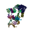
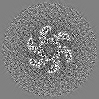
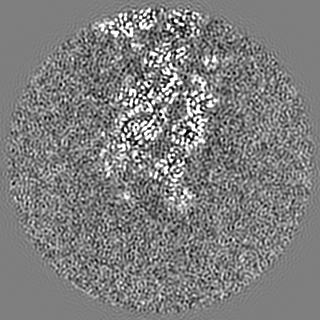
| Entry | Database: EMDB / ID: EMD-20644 | |||||||||||||||
|---|---|---|---|---|---|---|---|---|---|---|---|---|---|---|---|---|
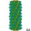
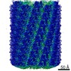
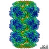
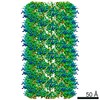
| Title | CryoEM Structure of Pyocin R2 - precontracted - collar | |||||||||||||||
 Map data Map data | Structure of Pyocin R2 precontracted collar | |||||||||||||||
 Sample Sample |
| |||||||||||||||
 Keywords Keywords | bacteriocin / pyocin / ANTIMICROBIAL PROTEIN | |||||||||||||||
| Function / homology |  Function and homology information Function and homology informationTail tube protein / Phage tail tube protein FII / : / Tail sheath protein Gp18 domain III N-terminal region / : / Tail sheath protein, subtilisin-like domain / Phage tail sheath protein subtilisin-like domain / Tail sheath protein, C-terminal domain / Phage tail sheath C-terminal domain Similarity search - Domain/homology | |||||||||||||||
| Biological species |  Pseudomonas aeruginosa PAO1 (bacteria) / Pseudomonas aeruginosa (strain ATCC 15692 / DSM 22644 / CIP 104116 / JCM 14847 / LMG 12228 / 1C / PRS 101 / PAO1) (bacteria) Pseudomonas aeruginosa PAO1 (bacteria) / Pseudomonas aeruginosa (strain ATCC 15692 / DSM 22644 / CIP 104116 / JCM 14847 / LMG 12228 / 1C / PRS 101 / PAO1) (bacteria) | |||||||||||||||
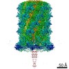
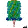
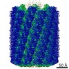
| Method | single particle reconstruction / cryo EM / Resolution: 3.8 Å | |||||||||||||||
 Authors Authors | Ge P / Avaylon J | |||||||||||||||
| Funding support |  United States, United States,  Switzerland, 4 items Switzerland, 4 items
| |||||||||||||||
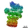
 Citation Citation | Journal: Nature / Year: 2020 Title: Action of a minimal contractile bactericidal nanomachine. Authors: Peng Ge / Dean Scholl / Nikolai S Prokhorov / Jaycob Avaylon / Mikhail M Shneider / Christopher Browning / Sergey A Buth / Michel Plattner / Urmi Chakraborty / Ke Ding / Petr G Leiman / Jeff ...Authors: Peng Ge / Dean Scholl / Nikolai S Prokhorov / Jaycob Avaylon / Mikhail M Shneider / Christopher Browning / Sergey A Buth / Michel Plattner / Urmi Chakraborty / Ke Ding / Petr G Leiman / Jeff F Miller / Z Hong Zhou /   Abstract: R-type bacteriocins are minimal contractile nanomachines that hold promise as precision antibiotics. Each bactericidal complex uses a collar to bridge a hollow tube with a contractile sheath loaded ...R-type bacteriocins are minimal contractile nanomachines that hold promise as precision antibiotics. Each bactericidal complex uses a collar to bridge a hollow tube with a contractile sheath loaded in a metastable state by a baseplate scaffold. Fine-tuning of such nucleic acid-free protein machines for precision medicine calls for an atomic description of the entire complex and contraction mechanism, which is not available from baseplate structures of the (DNA-containing) T4 bacteriophage. Here we report the atomic model of the complete R2 pyocin in its pre-contraction and post-contraction states, each containing 384 subunits of 11 unique atomic models of 10 gene products. Comparison of these structures suggests the following sequence of events during pyocin contraction: tail fibres trigger lateral dissociation of baseplate triplexes; the dissociation then initiates a cascade of events leading to sheath contraction; and this contraction converts chemical energy into mechanical force to drive the iron-tipped tube across the bacterial cell surface, killing the bacterium. | |||||||||||||||
| History |
|
- Structure visualization
| Movie |
Movie viewer |
|---|---|
| Structure viewer | EM map: SurfViewMolmilJmol/JSmol |
| Supplemental images |

- Downloads & links
-EMDB archive
| Map data | emd_20644.map.gz | 115.8 MB | EMDB map data format | |
|---|---|---|---|---|
| Header (meta data) | emd-20644-v30.xmlemd-20644.xml | 17 KB 17 KB | Display Display | EMDB header |
| FSC (resolution estimation) | emd_20644_fsc.xml | 11.4 KB | Display | FSC data file |
| Images | 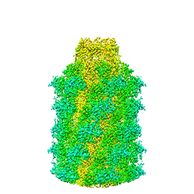 emd_20644.png emd_20644.png | 145.5 KB | ||
| Filedesc metadata | emd-20644.cif.gz | 6.1 KB | ||
| Archive directory |  http://ftp.pdbj.org/pub/emdb/structures/EMD-20644ftp://ftp.pdbj.org/pub/emdb/structures/EMD-20644 http://ftp.pdbj.org/pub/emdb/structures/EMD-20644ftp://ftp.pdbj.org/pub/emdb/structures/EMD-20644 | HTTPS FTP |
-Related structure data
| Related structure data |  6u5fMC  5cesC  6pytC 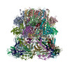 6u5bC 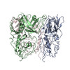 6u5hC 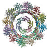 6u5jC 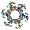 6u5kC C: citing same article ( M: atomic model generated by this map |
|---|---|
| Similar structure data |
-Links
| EMDB pages | EMDB (EBI/PDBe) / EMDataResource |
|---|
-Map
| File | Download / File: emd_20644.map.gz / Format: CCP4 / Size: 125 MB / Type: IMAGE STORED AS FLOATING POINT NUMBER (4 BYTES) | ||||||||||||||||||||||||||||||||||||||||||||||||||||||||||||||||||||
|---|---|---|---|---|---|---|---|---|---|---|---|---|---|---|---|---|---|---|---|---|---|---|---|---|---|---|---|---|---|---|---|---|---|---|---|---|---|---|---|---|---|---|---|---|---|---|---|---|---|---|---|---|---|---|---|---|---|---|---|---|---|---|---|---|---|---|---|---|---|
| Annotation | Structure of Pyocin R2 precontracted collar | ||||||||||||||||||||||||||||||||||||||||||||||||||||||||||||||||||||
| Projections & slices | Image control
Images are generated by Spider. | ||||||||||||||||||||||||||||||||||||||||||||||||||||||||||||||||||||
| Voxel size | X=Y=Z: 1.041 Å | ||||||||||||||||||||||||||||||||||||||||||||||||||||||||||||||||||||
| Density |
| ||||||||||||||||||||||||||||||||||||||||||||||||||||||||||||||||||||
| Symmetry | Space group: 1 | ||||||||||||||||||||||||||||||||||||||||||||||||||||||||||||||||||||
| Details | EMDB XML:
CCP4 map header:
| ||||||||||||||||||||||||||||||||||||||||||||||||||||||||||||||||||||
 Z (Sec.)
 Y (Row.)
 X (Col.)


-Supplemental data
- Sample components
-Entire : Pyocin R2
| Entire | Name: Pyocin R2 |
|---|---|
| Components |
|
-Supramolecule #1: Pyocin R2
| Supramolecule | Name: Pyocin R2 / type: complex / ID: 1 / Parent: 0 / Macromolecule list: all |
|---|---|
| Source (natural) | Organism: Pseudomonas aeruginosa PAO1 (bacteria) |
-Macromolecule #1: Collar PA0615
| Macromolecule | Name: Collar PA0615 / type: protein_or_peptide / ID: 1 / Number of copies: 6 / Enantiomer: LEVO |
|---|---|
| Source (natural) | Organism: Pseudomonas aeruginosa (strain ATCC 15692 / DSM 22644 / CIP 104116 / JCM 14847 / LMG 12228 / 1C / PRS 101 / PAO1) (bacteria) Strain: ATCC 15692 / DSM 22644 / CIP 104116 / JCM 14847 / LMG 12228 / 1C / PRS 101 / PAO1 |
| Molecular weight | Theoretical: 18.965461 KDa |
| Sequence | String: MPEQAVTLEA LYAAIEQVLR ERLPEAQLIG FWPGVPENTP AVSLEIAELL PERDPGTGES ALLCRLQARI MVPPGADRQA VSIACGIVR TLREQTWNLS LQPARFVRSA VDGSREELKS LRVWLVEWTQ SLRLGDPEWA WEDQPPGSLM LGFDPQTGPG H EPDYFAPE ALA UniProtKB: Phage protein |
-Macromolecule #2: Sheath PA0622
| Macromolecule | Name: Sheath PA0622 / type: protein_or_peptide / ID: 2 / Number of copies: 24 / Enantiomer: LEVO |
|---|---|
| Source (natural) | Organism: Pseudomonas aeruginosa (strain ATCC 15692 / DSM 22644 / CIP 104116 / JCM 14847 / LMG 12228 / 1C / PRS 101 / PAO1) (bacteria) Strain: ATCC 15692 / DSM 22644 / CIP 104116 / JCM 14847 / LMG 12228 / 1C / PRS 101 / PAO1 |
| Molecular weight | Theoretical: 41.247332 KDa |
| Sequence | String: MSFFHGVTVT NVDIGARTIA LPASSVIGLC DVFTPGAQAS AKPNVPVLLT SKKDAAAAFG IGSSIYLACE AIYNRAQAVI VAVGVETAE TPEAQASAVI GGISAAGERT GLQALLDGKS RFNAQPRLLV APGHSAQQAV ATAMDGLAEK LRAIAILDGP N STDEAAVA ...String: MSFFHGVTVT NVDIGARTIA LPASSVIGLC DVFTPGAQAS AKPNVPVLLT SKKDAAAAFG IGSSIYLACE AIYNRAQAVI VAVGVETAE TPEAQASAVI GGISAAGERT GLQALLDGKS RFNAQPRLLV APGHSAQQAV ATAMDGLAEK LRAIAILDGP N STDEAAVA YAKNFGSKRL FMVDPGVQVW DSATNAARNA PASAYAAGLF AWTDAEYGFW SSPSNKEIKG VTGTSRPVEF LD GDETCRA NLLNNANIAT IIRDDGYRLW GNRTLSSDSK WAFVTRVRTM DLVMDAILAG HKWAVDRGIT KTYVKDVTEG LRA FMRDLK NQGAVINFEV YADPDLNSAS QLAQGKVYWN IRFTDVPPAE NPNFRVEVTD QWLTEVLDVA UniProtKB: Probable bacteriophage protein |
-Macromolecule #3: Tube PA0623
| Macromolecule | Name: Tube PA0623 / type: protein_or_peptide / ID: 3 / Number of copies: 24 / Enantiomer: LEVO |
|---|---|
| Source (natural) | Organism: Pseudomonas aeruginosa (strain ATCC 15692 / DSM 22644 / CIP 104116 / JCM 14847 / LMG 12228 / 1C / PRS 101 / PAO1) (bacteria) Strain: ATCC 15692 / DSM 22644 / CIP 104116 / JCM 14847 / LMG 12228 / 1C / PRS 101 / PAO1 |
| Molecular weight | Theoretical: 17.957352 KDa |
| Sequence | String: MIPQTLTNTN LFIDGVSFAG DVPSLTLPKL AVKTEQYRAG GMDAPVSIDM GLEAMEAKFS TNGARREALN FFGLADQSAF NGVFRGSFK GQKGASVPVV ATLRGLLKEV DPGDWKAGEK AEFKYAVAVS YYKLEVDGRE VYEIDPVNGV RAINGVDQLA G MRNDLGL UniProtKB: Probable bacteriophage protein |
-Experimental details
-Structure determination
| Method | cryo EM |
|---|---|
 Processing Processing | single particle reconstruction |
| Aggregation state | particle |
-Sample preparation
| Buffer | pH: 7.4 Component:
| |||||||||
|---|---|---|---|---|---|---|---|---|---|---|
| Grid | Support film - Material: CARBON / Support film - topology: HOLEY ARRAY / Details: unspecified | |||||||||
| Vitrification | Cryogen name: ETHANE / Chamber humidity: 100 % / Chamber temperature: 295 K / Instrument: FEI VITROBOT MARK IV |
- Electron microscopy
| Microscope | FEI TITAN KRIOS |
|---|---|
| Temperature | Min: 80.0 K / Max: 81.0 K |
| Specialist optics | Energy filter - Name: GIF Quantum LS / Energy filter - Lower energy threshold: -10 eV / Energy filter - Upper energy threshold: 10 eV |
| Image recording | Film or detector model: GATAN K2 QUANTUM (4k x 4k) / Detector mode: COUNTING / Digitization - Frames/image: 3-20 / Number grids imaged: 1 / Number real images: 7331 / Average exposure time: 10.0 sec. / Average electron dose: 80.0 e/Å2 |
| Electron beam | Acceleration voltage: 300 kV / Electron source:  FIELD EMISSION GUN FIELD EMISSION GUN |
| Electron optics | C2 aperture diameter: 70.0 µm / Calibrated defocus max: 3.4 µm / Calibrated defocus min: 1.1 µm / Illumination mode: FLOOD BEAM / Imaging mode: BRIGHT FIELD / Cs: 2.7 mm / Nominal defocus max: 2.16 µm / Nominal defocus min: 2.16 µm / Nominal magnification: 130000 |
| Sample stage | Specimen holder model: FEI TITAN KRIOS AUTOGRID HOLDER / Cooling holder cryogen: NITROGEN |
| Experimental equipment |  Model: Titan Krios / Image courtesy: FEI Company |
+Image processing

-Atomic model buiding 1
| Refinement | Space: REAL / Protocol: AB INITIO MODEL |
|---|---|
| Output model | PDB-6u5f: |
