 Movie
Movie Controller

[English] 日本語
 Yorodumi
- EMDB-23497: Filamentous actin decorated with human cardiac myosin binding pro... -
+ Open data

- Basic information
| Entry | Database: EMDB / ID: EMD-23497 | ||||||||||||
|---|---|---|---|---|---|---|---|---|---|---|---|---|---|
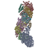
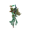
| Title | Filamentous actin decorated with human cardiac myosin binding protein C C2 domain | ||||||||||||
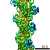
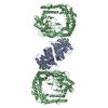
 Map data Map data | Filamentous actin decorated with human cardiac myosin binding protein C C2 domain | ||||||||||||
 Sample Sample |
| ||||||||||||
 Keywords Keywords | myosin binding protein C / actin / thin filament / MOTOR PROTEIN | ||||||||||||
| Function / homology |  Function and homology information Function and homology informationC zone / regulation of muscle filament sliding / striated muscle myosin thick filament / regulation of striated muscle contraction / cardiac myofibril / actin-myosin filament sliding / A band / Striated Muscle Contraction / regulation of cardiac muscle cell contraction / M band ...C zone / regulation of muscle filament sliding / striated muscle myosin thick filament / regulation of striated muscle contraction / cardiac myofibril / actin-myosin filament sliding / A band / Striated Muscle Contraction / regulation of cardiac muscle cell contraction / M band / structural constituent of muscle / sarcomere organization / ventricular cardiac muscle tissue morphogenesis / myosin heavy chain binding / myosin binding / heart contraction / mesenchyme migration / ATPase activator activity / heart morphogenesis / cardiac muscle contraction / titin binding / actin filament organization / sarcomere / filopodium / actin filament / lamellipodium / actin cytoskeleton / actin binding / cell body / cell adhesion / positive regulation of gene expression / ATP binding / metal ion binding / identical protein binding / cytoplasm / cytosol Similarity search - Function | ||||||||||||
| Biological species |   Homo sapiens (human) Homo sapiens (human) | ||||||||||||
| Method | helical reconstruction / cryo EM / Resolution: 6.1 Å | ||||||||||||
 Authors Authors | Galkin VE | ||||||||||||
| Funding support |  United States, 3 items United States, 3 items
| ||||||||||||
 Citation Citation | Journal: J Mol Biol / Year: 2021 Title: Interaction of the C2 Ig-like Domain of Cardiac Myosin Binding Protein-C with F-actin. Authors: Cristina M Risi / Malay Patra / Betty Belknap / Samantha P Harris / Howard D White / Vitold E Galkin / Abstract: Cardiac muscle contraction depends on interactions between thick (myosin) and thin (actin) filaments (TFs). TFs are regulated by intracellular Ca levels. Under activating conditions Ca binds to the ...Cardiac muscle contraction depends on interactions between thick (myosin) and thin (actin) filaments (TFs). TFs are regulated by intracellular Ca levels. Under activating conditions Ca binds to the troponin complex and displaces tropomyosin from myosin binding sites on the TF surface to allow actomyosin interactions. Recent studies have shown that in addition to Ca, the first four N-terminal domains (NTDs) of cardiac myosin binding protein C (cMyBP-C) (e.g. C0, C1, M and C2), are potent modulators of the TF activity, but the mechanism of their collective action is poorly understood. Previously, we showed that C1 activates the TF at low Ca and C0 stabilizes binding of C1 to the TF, but the ability of C2 to bind and/or affect the TF remains unknown. Here we obtained 7.5 Å resolution cryo-EM reconstruction of C2-decorated actin filaments to demonstrate that C2 binds to actin in a single structural mode that does not activate the TF unlike the polymorphic binding of C0 and C1 to actin. Comparison of amino acid sequences of C2 with either C0 or C1 shows low levels of identity between the residues involved in interactions with the TF but high levels of conservation for residues involved in Ig fold stabilization. This provides a structural basis for strikingly different interactions of structurally homologous C0, C1 and C2 with the TF. Our detailed analysis of the interaction of C2 with the actin filament provides crucial information required to model the collective action of cMyBP-C NTDs on the cardiac TF. | ||||||||||||
| History |
|
- Structure visualization
| Movie |
Movie viewer |
|---|---|
| Structure viewer | EM map: SurfViewMolmilJmol/JSmol |
| Supplemental images |

- Downloads & links
-EMDB archive
| Map data | emd_23497.map.gz | 10.1 MB | EMDB map data format | |
|---|---|---|---|---|
| Header (meta data) | emd-23497-v30.xmlemd-23497.xml | 14 KB 14 KB | Display Display | EMDB header |
| Images |  emd_23497.png emd_23497.png | 269.2 KB | ||
| Filedesc metadata | emd-23497.cif.gz | 6 KB | ||
| Archive directory |  http://ftp.pdbj.org/pub/emdb/structures/EMD-23497ftp://ftp.pdbj.org/pub/emdb/structures/EMD-23497 http://ftp.pdbj.org/pub/emdb/structures/EMD-23497ftp://ftp.pdbj.org/pub/emdb/structures/EMD-23497 | HTTPS FTP |
-Related structure data
| Related structure data |  7lrgMC M: atomic model generated by this map C: citing same article ( |
|---|---|
| Similar structure data |


-Links
| EMDB pages | EMDB (EBI/PDBe) / EMDataResource |
|---|---|
| Related items in Molecule of the Month |


-Map
| File | Download / File: emd_23497.map.gz / Format: CCP4 / Size: 11.6 MB / Type: IMAGE STORED AS FLOATING POINT NUMBER (4 BYTES) | ||||||||||||||||||||||||||||||||||||||||||||||||||||||||||||||||||||
|---|---|---|---|---|---|---|---|---|---|---|---|---|---|---|---|---|---|---|---|---|---|---|---|---|---|---|---|---|---|---|---|---|---|---|---|---|---|---|---|---|---|---|---|---|---|---|---|---|---|---|---|---|---|---|---|---|---|---|---|---|---|---|---|---|---|---|---|---|---|
| Annotation | Filamentous actin decorated with human cardiac myosin binding protein C C2 domain | ||||||||||||||||||||||||||||||||||||||||||||||||||||||||||||||||||||
| Projections & slices | Image control
Images are generated by Spider. generated in cubic-lattice coordinate | ||||||||||||||||||||||||||||||||||||||||||||||||||||||||||||||||||||
| Voxel size | X=Y=Z: 1.08 Å | ||||||||||||||||||||||||||||||||||||||||||||||||||||||||||||||||||||
| Density |
| ||||||||||||||||||||||||||||||||||||||||||||||||||||||||||||||||||||
| Symmetry | Space group: 1 | ||||||||||||||||||||||||||||||||||||||||||||||||||||||||||||||||||||
| Details | EMDB XML:
CCP4 map header:
| ||||||||||||||||||||||||||||||||||||||||||||||||||||||||||||||||||||
 Z (Sec.)
 Y (Row.)
 X (Col.)


-Supplemental data
- Sample components
-Entire : Filamentous actin decorated with human cardiac myosin binding pro...
| Entire | Name: Filamentous actin decorated with human cardiac myosin binding protein C C2 domain |
|---|---|
| Components |
|
-Supramolecule #1: Filamentous actin decorated with human cardiac myosin binding pro...
| Supramolecule | Name: Filamentous actin decorated with human cardiac myosin binding protein C C2 domain type: complex / ID: 1 / Parent: 0 / Macromolecule list: all |
|---|
-Supramolecule #2: Filamentous actin
| Supramolecule | Name: Filamentous actin / type: complex / ID: 2 / Parent: 1 / Macromolecule list: #1 |
|---|---|
| Source (natural) | Organism: |
-Supramolecule #3: Cardiac myosin binding protein C C2 domain
| Supramolecule | Name: Cardiac myosin binding protein C C2 domain / type: complex / ID: 3 / Parent: 1 / Macromolecule list: #2 |
|---|---|
| Source (natural) | Organism: Homo sapiens (human) / Organ: heart |
-Macromolecule #1: Actin, alpha cardiac muscle 1
| Macromolecule | Name: Actin, alpha cardiac muscle 1 / type: protein_or_peptide / ID: 1 / Number of copies: 6 / Enantiomer: LEVO |
|---|---|
| Source (natural) | Organism: |
| Molecular weight | Theoretical: 42.064891 KDa |
| Sequence | String: MCDDEETTAL VCDNGSGLVK AGFAGDDAPR AVFPSIVGRP RHQGVMVGMG QKDSYVGDEA QSKRGILTLK YPIEHGIITN WDDMEKIWH HTFYNELRVA PEEHPTLLTE APLNPKANRE KMTQIMFETF NVPAMYVAIQ AVLSLYASGR TTGIVLDSGD G VTHNVPIY ...String: MCDDEETTAL VCDNGSGLVK AGFAGDDAPR AVFPSIVGRP RHQGVMVGMG QKDSYVGDEA QSKRGILTLK YPIEHGIITN WDDMEKIWH HTFYNELRVA PEEHPTLLTE APLNPKANRE KMTQIMFETF NVPAMYVAIQ AVLSLYASGR TTGIVLDSGD G VTHNVPIY EGYALPHAIM RLDLAGRDLT DYLMKILTER GYSFVTTAER EIVRDIKEKL CYVALDFENE MATAASSSSL EK SYELPDG QVITIGNERF RCPETLFQPS FIGMESAGIH ETTYNSIMKC DIDIRKDLYA NNVLSGGTTM YPGIADRMQK EIT ALAPST MKIKIIAPPE RKYSVWIGGS ILASLSTFQQ MWISKQEYDE AGPSIVHRKC F UniProtKB: Actin alpha cardiac muscle 1 |
-Macromolecule #2: Myosin-binding protein C, cardiac-type
| Macromolecule | Name: Myosin-binding protein C, cardiac-type / type: protein_or_peptide / ID: 2 / Number of copies: 6 / Enantiomer: LEVO |
|---|---|
| Source (natural) | Organism: Homo sapiens (human) |
| Molecular weight | Theoretical: 10.499937 KDa |
| Recombinant expression | Organism:  |
| Sequence | String: DEKKSTAFQK KLEPAYQVSK GHKIRLTVEL ADHDAEVKWL KNGQEIQMSG SKYIFESIGA KRTLTISQCS LADDAAYQCV VGGEKCSTE LFVKE UniProtKB: Myosin-binding protein C, cardiac-type |
-Experimental details
-Structure determination
| Method | cryo EM |
|---|---|
 Processing Processing | helical reconstruction |
| Aggregation state | helical array |
-Sample preparation
| Buffer | pH: 7 |
|---|---|
| Grid | Model: PELCO Ultrathin Carbon with Lacey Carbon / Material: COPPER / Mesh: 300 |
| Vitrification | Cryogen name: ETHANE / Chamber humidity: 100 % / Chamber temperature: 10 K / Instrument: FEI VITROBOT MARK IV |
- Electron microscopy
| Microscope | FEI TITAN KRIOS |
|---|---|
| Image recording | Film or detector model: GATAN K3 (6k x 4k) / Average electron dose: 35.0 e/Å2 Details: Images collected in movie-mode with 44 subframes at 0.85e-2/A per frame |
| Electron beam | Acceleration voltage: 300 kV / Electron source:  FIELD EMISSION GUN FIELD EMISSION GUN |
| Electron optics | Illumination mode: FLOOD BEAM / Imaging mode: BRIGHT FIELD |
| Sample stage | Specimen holder model: FEI TITAN KRIOS AUTOGRID HOLDER / Cooling holder cryogen: NITROGEN |
| Experimental equipment |  Model: Titan Krios / Image courtesy: FEI Company |
-Image processing
| Final reconstruction | Applied symmetry - Helical parameters - Δz: 27.3 Å Applied symmetry - Helical parameters - Δ&Phi: -166.5 ° Applied symmetry - Helical parameters - Axial symmetry: C1 (asymmetric) Resolution.type: BY AUTHOR / Resolution: 6.1 Å / Resolution method: FSC 0.143 CUT-OFF / Software - Name: IHRSR / Number images used: 43047 |
|---|---|
| Startup model | Type of model: OTHER / Details: randomized azimuthal angles used |
| Final angle assignment | Type: NOT APPLICABLE / Software - Name: SPIDER |


